 Movie
Movie Controller


+ Open data

- Basic information
| Entry | Database: PDB / ID: 4fu6 | ||||||
|---|---|---|---|---|---|---|---|
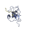
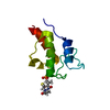
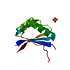
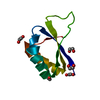
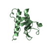
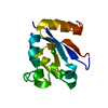
| Title | Crystal structure of the PSIP1 PWWP domain | ||||||
 Components Components | PC4 and SFRS1-interacting protein | ||||||
 Keywords Keywords |  TRANSCRIPTION / Structural Genomics Consortium / SGC TRANSCRIPTION / Structural Genomics Consortium / SGC | ||||||
| Function / homology |  Function and homology informationsupercoiled DNA binding / Integration of viral DNA into host genomic DNA / Autointegration results in viral DNA circles / Formation of WDR5-containing histone-modifying complexes / 2-LTR circle formation / Vpr-mediated nuclear import of PICs / Integration of provirus / APOBEC3G mediated resistance to HIV-1 infection / mRNA 5'-splice site recognition / heterochromatin ...supercoiled DNA binding / Integration of viral DNA into host genomic DNA / Autointegration results in viral DNA circles / Formation of WDR5-containing histone-modifying complexes / 2-LTR circle formation / Vpr-mediated nuclear import of PICs / Integration of provirus / APOBEC3G mediated resistance to HIV-1 infection / mRNA 5'-splice site recognition / heterochromatin / nuclear periphery / euchromatin / response to heat / DNA-binding transcription factor binding / response to oxidative stress / transcription coactivator activity / chromatin remodeling / chromatin binding / positive regulation of transcription by RNA polymerase II / RNA binding / nucleoplasm / nucleus / cytosol Function and homology informationsupercoiled DNA binding / Integration of viral DNA into host genomic DNA / Autointegration results in viral DNA circles / Formation of WDR5-containing histone-modifying complexes / 2-LTR circle formation / Vpr-mediated nuclear import of PICs / Integration of provirus / APOBEC3G mediated resistance to HIV-1 infection / mRNA 5'-splice site recognition / heterochromatin ...supercoiled DNA binding / Integration of viral DNA into host genomic DNA / Autointegration results in viral DNA circles / Formation of WDR5-containing histone-modifying complexes / 2-LTR circle formation / Vpr-mediated nuclear import of PICs / Integration of provirus / APOBEC3G mediated resistance to HIV-1 infection / mRNA 5'-splice site recognition / heterochromatin / nuclear periphery / euchromatin / response to heat / DNA-binding transcription factor binding / response to oxidative stress / transcription coactivator activity / chromatin remodeling / chromatin binding / positive regulation of transcription by RNA polymerase II / RNA binding / nucleoplasm / nucleus / cytosolSimilarity search - Function | ||||||
| Biological species |  Homo sapiens (human) Homo sapiens (human) | ||||||
| Method | X-RAY DIFFRACTION / SYNCHROTRON / MOLECULAR REPLACEMENT / molecular replacement / Resolution: 2.1 Å | ||||||
 Authors Authors | Qin, S. / Tempel, W. / Xu, C. / Wu, H. / Dong, A. / Cerovina, T. / Bountra, C. / Arrowsmith, C.H. / Edwards, A.M. / Min, J. / Structural Genomics Consortium (SGC) | ||||||
 Citation Citation | Journal: Trends Biochem.Sci. / Year: 2014 Title: Structure and function of the nucleosome-binding PWWP domain. Authors: Qin, S. / Min, J. | ||||||
| History |
|
- Structure visualization
| Structure viewer | Molecule: MolmilJmol/JSmol |
|---|
- Downloads & links
-Download
| PDBx/mmCIF format | 4fu6.cif.gz | 56.8 KB | Display | PDBx/mmCIF format |
|---|---|---|---|---|
| PDB format | pdb4fu6.ent.gz | 39.3 KB | Display | PDB format |
| PDBx/mmJSON format | 4fu6.json.gz | Tree view | PDBx/mmJSON format | |
| Others |  Other downloads Other downloads |
-Validation report
| Arichive directory | https://data.pdbj.org/pub/pdb/validation_reports/fu/4fu6ftp://data.pdbj.org/pub/pdb/validation_reports/fu/4fu6 | HTTPS FTP |
|---|
-Related structure data
| Related structure data |  3qbyS S: Starting model for refinement |
|---|---|
| Similar structure data |


-Links
 PDBj
PDBj


- Assembly
| Deposited unit | 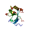
| |||||||||
|---|---|---|---|---|---|---|---|---|---|---|
| 1 |
| |||||||||
| Unit cell |
| |||||||||
| Components on special symmetry positions |
|
-Components
| #1: Protein | Mass: 17518.379 Da / Num. of mol.: 1 / Fragment: UNP residues 1-135 Source method: isolated from a genetically manipulated source Source: (gene. exp.) Homo sapiens (human) / Gene: PSIP1, DFS70, LEDGF, PSIP2 / Plasmid: pET28-MHL / Production host:  Escherichia coli (E. coli) / Strain (production host): BL21(DE3) / References: UniProt: O75475 Escherichia coli (E. coli) / Strain (production host): BL21(DE3) / References: UniProt: O75475 | ||||||||
|---|---|---|---|---|---|---|---|---|---|
| #2: Chemical | Sulfate  Mass: 96.063 Da / Num. of mol.: 3 / Source method: obtained synthetically / Formula: SO4 Mass: 96.063 Da / Num. of mol.: 3 / Source method: obtained synthetically / Formula: SO4#3: Chemical | ChemComp-GOL / | Glycerol  Mass: 92.094 Da / Num. of mol.: 1 / Source method: obtained synthetically / Formula: C3H8O3 Mass: 92.094 Da / Num. of mol.: 1 / Source method: obtained synthetically / Formula: C3H8O3#4: Chemical | ChemComp-UNX /   Num. of mol.: 5 / Source method: obtained synthetically Num. of mol.: 5 / Source method: obtained synthetically#5: Water | ChemComp-HOH / | Water Mass: 18.015 Da / Num. of mol.: 30 / Source method: isolated from a natural source / Formula: H2O Mass: 18.015 Da / Num. of mol.: 30 / Source method: isolated from a natural source / Formula: H2OSequence details | CALCULATED VALUES FOR MATTHEWS COEFFICIENT AND SOLVENT CONTENT(2.35/47.77) ARE BASED ON THE ENTIRE ...CALCULATED | |
-Experimental details
-Experiment
| Experiment | Method: X-RAY DIFFRACTION / Number of used crystals: 1 |
|---|
- Sample preparation
| Crystal | Density Matthews: 2.35 Å3/Da / Density % sol: 47.77 % |
|---|---|
| Crystal grow | Temperature: 291 K / Method: vapor diffusion / pH: 8.5 Details: The protein sample was treated with V8 protease. 2.5M ammonium sulfate, 0.1M TRIS, pH 8.5, vapor diffusion, temperature 291K |
-Data collection
| Diffraction | Mean temperature: 100 K | |||||||||||||||||||||||||||||||||||||||||||||||||||||||||||||||||||||||||||||||||||||||||||||||||||||||||||||||||||||||||||||||||||||||||||||||||||
|---|---|---|---|---|---|---|---|---|---|---|---|---|---|---|---|---|---|---|---|---|---|---|---|---|---|---|---|---|---|---|---|---|---|---|---|---|---|---|---|---|---|---|---|---|---|---|---|---|---|---|---|---|---|---|---|---|---|---|---|---|---|---|---|---|---|---|---|---|---|---|---|---|---|---|---|---|---|---|---|---|---|---|---|---|---|---|---|---|---|---|---|---|---|---|---|---|---|---|---|---|---|---|---|---|---|---|---|---|---|---|---|---|---|---|---|---|---|---|---|---|---|---|---|---|---|---|---|---|---|---|---|---|---|---|---|---|---|---|---|---|---|---|---|---|---|---|---|---|
| Diffraction source | Source: SYNCHROTRON / Site: APS  / Beamline: 23-ID-D / Wavelength: 0.97944 Å / Beamline: 23-ID-D / Wavelength: 0.97944 Å | |||||||||||||||||||||||||||||||||||||||||||||||||||||||||||||||||||||||||||||||||||||||||||||||||||||||||||||||||||||||||||||||||||||||||||||||||||
| Detector | Type: MAR scanner 300 mm plate / Detector: IMAGE PLATE / Date: Jun 22, 2012 | |||||||||||||||||||||||||||||||||||||||||||||||||||||||||||||||||||||||||||||||||||||||||||||||||||||||||||||||||||||||||||||||||||||||||||||||||||
| Radiation | Protocol: SINGLE WAVELENGTH / Monochromatic (M) / Laue (L): M / Scattering type: x-ray | |||||||||||||||||||||||||||||||||||||||||||||||||||||||||||||||||||||||||||||||||||||||||||||||||||||||||||||||||||||||||||||||||||||||||||||||||||
| Radiation wavelength | Wavelength: 0.97944 Å / Relative weight: 1 | |||||||||||||||||||||||||||||||||||||||||||||||||||||||||||||||||||||||||||||||||||||||||||||||||||||||||||||||||||||||||||||||||||||||||||||||||||
| Reflection | Resolution: 2.1→50 Å / Num. obs: 10584 / % possible obs: 100 % / Redundancy: 13.4 % / Rmerge(I) obs: 0.081 / Χ2: 1.763 / Net I/σ(I): 10.2 | |||||||||||||||||||||||||||||||||||||||||||||||||||||||||||||||||||||||||||||||||||||||||||||||||||||||||||||||||||||||||||||||||||||||||||||||||||
| Reflection shell |
|
-Phasing
| Phasing | Method: molecular replacement |
|---|
- Processing
| Software |
| |||||||||||||||||||||||||||||||||||||||||||||||||||||||||||||||||||||||||||||||||||||||||||||||||||||||||||||||||||||||||||||||||||||||||||||||||||
|---|---|---|---|---|---|---|---|---|---|---|---|---|---|---|---|---|---|---|---|---|---|---|---|---|---|---|---|---|---|---|---|---|---|---|---|---|---|---|---|---|---|---|---|---|---|---|---|---|---|---|---|---|---|---|---|---|---|---|---|---|---|---|---|---|---|---|---|---|---|---|---|---|---|---|---|---|---|---|---|---|---|---|---|---|---|---|---|---|---|---|---|---|---|---|---|---|---|---|---|---|---|---|---|---|---|---|---|---|---|---|---|---|---|---|---|---|---|---|---|---|---|---|---|---|---|---|---|---|---|---|---|---|---|---|---|---|---|---|---|---|---|---|---|---|---|---|---|---|
| Refinement | Method to determine structure: MOLECULAR REPLACEMENT Starting model: pdb entry 3QBY Resolution: 2.1→40 Å / Cor.coef. Fo:Fc: 0.957 / Cor.coef. Fo:Fc free: 0.942 / WRfactor Rfree: 0.228 / WRfactor Rwork: 0.198 / Occupancy max: 1 / Occupancy min: 0.5 / SU B: 7.875 / SU ML: 0.103 / Cross valid method: THROUGHOUT / σ(F): 0 / ESU R: 0.148 / ESU R Free: 0.141 / Stereochemistry target values: MAXIMUM LIKELIHOOD Details: HYDROGENS HAVE BEEN ADDED IN THE RIDING POSITIONS U VALUES : WITH TLS ADDED. the program COOT and the molprobity server were also used. CAVEAT: the C-terminal direction of the protein main ...Details: HYDROGENS HAVE BEEN ADDED IN THE RIDING POSITIONS U VALUES : WITH TLS ADDED. the program COOT and the molprobity server were also used. CAVEAT: the C-terminal direction of the protein main chain at LYS-91 is not resolved by electron density.
| |||||||||||||||||||||||||||||||||||||||||||||||||||||||||||||||||||||||||||||||||||||||||||||||||||||||||||||||||||||||||||||||||||||||||||||||||||
| Solvent computation | Ion probe radii: 0.8 Å / Shrinkage radii: 0.8 Å / VDW probe radii: 1.2 Å / Solvent model: MASK BULK SOLVENT | |||||||||||||||||||||||||||||||||||||||||||||||||||||||||||||||||||||||||||||||||||||||||||||||||||||||||||||||||||||||||||||||||||||||||||||||||||
| Displacement parameters | Biso max: 107.07 Å2 / Biso mean: 46.012 Å2 / Biso min: 27.01 Å2
| |||||||||||||||||||||||||||||||||||||||||||||||||||||||||||||||||||||||||||||||||||||||||||||||||||||||||||||||||||||||||||||||||||||||||||||||||||
| Refinement step | Cycle: LAST / Resolution: 2.1→40 Å
| |||||||||||||||||||||||||||||||||||||||||||||||||||||||||||||||||||||||||||||||||||||||||||||||||||||||||||||||||||||||||||||||||||||||||||||||||||
| Refine LS restraints |
| |||||||||||||||||||||||||||||||||||||||||||||||||||||||||||||||||||||||||||||||||||||||||||||||||||||||||||||||||||||||||||||||||||||||||||||||||||
| LS refinement shell | Refine-ID: X-RAY DIFFRACTION / Total num. of bins used: 20
| |||||||||||||||||||||||||||||||||||||||||||||||||||||||||||||||||||||||||||||||||||||||||||||||||||||||||||||||||||||||||||||||||||||||||||||||||||
| Refinement TLS params. | Method: refined / Origin x: 7.1569 Å / Origin y: 5.8533 Å / Origin z: 52.1674 Å
| |||||||||||||||||||||||||||||||||||||||||||||||||||||||||||||||||||||||||||||||||||||||||||||||||||||||||||||||||||||||||||||||||||||||||||||||||||
| Refinement TLS group |
|
