 Movie
Movie Controller

[English] 日本語
 Yorodumi
- PDB-3r9l: Crystal structure of nucleoside diphosphate kinase from Giardia l... -
+ Open data

- Basic information
| Entry | Database: PDB / ID: 3r9l | ||||||
|---|---|---|---|---|---|---|---|
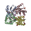
| Title | Crystal structure of nucleoside diphosphate kinase from Giardia lamblia featuring a disordered dinucleotide binding site | ||||||
 Components Components | Nucleoside diphosphate kinase Nucleoside-diphosphate kinase Nucleoside-diphosphate kinase | ||||||
 Keywords Keywords | TRANSFERASE / Structural Genomics / Seattle Structural Genomics Center for Infectious Disease / SSGCID / giardiasis / intestinal disease / protozoan parasite / water contaminant / kinase / phosphoryltransfer / ADP | ||||||
| Function / homology |  Function and homology informationnucleoside-diphosphate kinase / CTP biosynthetic process / UTP biosynthetic process / GTP biosynthetic process / nucleoside diphosphate kinase activity Function and homology informationnucleoside-diphosphate kinase / CTP biosynthetic process / UTP biosynthetic process / GTP biosynthetic process / nucleoside diphosphate kinase activitySimilarity search - Function | ||||||
| Biological species |   Giardia lamblia (eukaryote) Giardia lamblia (eukaryote) | ||||||
| Method | X-RAY DIFFRACTION / SYNCHROTRON / MOLECULAR REPLACEMENT / molecular replacement / Resolution: 2.65 Å | ||||||
 Authors Authors | Seattle Structural Genomics Center for Infectious Disease (SSGCID) | ||||||
 Citation Citation | Journal: To be Published Title: Crystal structure of nucleoside diphosphate kinase from Giardia lamblia featuring a disordered dinucleotide binding site Authors: Edwards, T.E. / Gardberg, A.S. / Sankaran, B. / Seattle Structural Genomics Center for Infectious Disease (SSGCID) | ||||||
| History |
|
- Structure visualization
| Structure viewer | Molecule: MolmilJmol/JSmol |
|---|
- Downloads & links
-Download
| PDBx/mmCIF format | 3r9l.cif.gz | 58.7 KB | Display | PDBx/mmCIF format |
|---|---|---|---|---|
| PDB format | pdb3r9l.ent.gz | 41.8 KB | Display | PDB format |
| PDBx/mmJSON format | 3r9l.json.gz | Tree view | PDBx/mmJSON format | |
| Others |  Other downloads Other downloads |
-Validation report
| Arichive directory | https://data.pdbj.org/pub/pdb/validation_reports/r9/3r9lftp://data.pdbj.org/pub/pdb/validation_reports/r9/3r9l | HTTPS FTP |
|---|
-Related structure data
| Related structure data |  1npkS S: Starting model for refinement |
|---|---|
| Similar structure data | |
| Other databases |

-Links
 PDBj
PDBj- Assembly
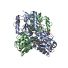
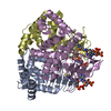
| Deposited unit | 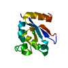
| ||||||||
|---|---|---|---|---|---|---|---|---|---|
| 1 | x 6
| ||||||||
| Unit cell |
|
-Components
| #1: Protein | Nucleoside-diphosphate kinase Mass: 17114.713 Da / Num. of mol.: 1 Source method: isolated from a genetically manipulated source Source: (gene. exp.) Giardia lamblia (eukaryote) / Strain: ATCC 50803 / WB clone C6 / Gene: GL50803_11301 / Production host:  Escherichia coli (E. coli) / References: UniProt: A8BMG9, nucleoside-diphosphate kinase Escherichia coli (E. coli) / References: UniProt: A8BMG9, nucleoside-diphosphate kinase |
|---|---|
| #2: Chemical | ChemComp-CL / Chloride  Mass: 35.453 Da / Num. of mol.: 1 / Source method: obtained synthetically / Formula: Cl Mass: 35.453 Da / Num. of mol.: 1 / Source method: obtained synthetically / Formula: Cl |
| #3: Water | ChemComp-HOH / Water Mass: 18.015 Da / Num. of mol.: 13 / Source method: isolated from a natural source / Formula: H2O Mass: 18.015 Da / Num. of mol.: 13 / Source method: isolated from a natural source / Formula: H2O |
-Experimental details
-Experiment
| Experiment | Method: X-RAY DIFFRACTION / Number of used crystals: 1 |
|---|
- Sample preparation
| Crystal | Density Matthews: 3.62 Å3/Da / Density % sol: 65.99 % |
|---|---|
| Crystal grow | Temperature: 289 K / Method: vapor diffusion, sitting drop / pH: 7.9 Details: GilaA.00438.a.A1 PW27083 at 24.97 mg/mL in 25 mM Hepes pH 7.0, 0.5 M NaCl, 5% glycerol, 2 mM DTT, 0.025% azide against CSHT C11 focus screen 0.1 M Hepes pH 7.0, 0.9 M Na phosphate, 0.9 M K ...Details: GilaA.00438.a.A1 PW27083 at 24.97 mg/mL in 25 mM Hepes pH 7.0, 0.5 M NaCl, 5% glycerol, 2 mM DTT, 0.025% azide against CSHT C11 focus screen 0.1 M Hepes pH 7.0, 0.9 M Na phosphate, 0.9 M K phosphate with 25% ethylene glycol as cryo-protectant, crystal tracking ID 216300h9, VAPOR DIFFUSION, SITTING DROP, temperature 289K |
-Data collection
| Diffraction | Mean temperature: 100 K | |||||||||||||||||||||||||||||||||||||||||||||||||||||||||||||||||||||||||||||||||||||||||||||||||||||||||||||||||||||||||||||||||||||||||||||||||||||||||||||||||||||||||||||||||||||||||||||
|---|---|---|---|---|---|---|---|---|---|---|---|---|---|---|---|---|---|---|---|---|---|---|---|---|---|---|---|---|---|---|---|---|---|---|---|---|---|---|---|---|---|---|---|---|---|---|---|---|---|---|---|---|---|---|---|---|---|---|---|---|---|---|---|---|---|---|---|---|---|---|---|---|---|---|---|---|---|---|---|---|---|---|---|---|---|---|---|---|---|---|---|---|---|---|---|---|---|---|---|---|---|---|---|---|---|---|---|---|---|---|---|---|---|---|---|---|---|---|---|---|---|---|---|---|---|---|---|---|---|---|---|---|---|---|---|---|---|---|---|---|---|---|---|---|---|---|---|---|---|---|---|---|---|---|---|---|---|---|---|---|---|---|---|---|---|---|---|---|---|---|---|---|---|---|---|---|---|---|---|---|---|---|---|---|---|---|---|---|---|---|
| Diffraction source | Source: SYNCHROTRON / Site: ALS  / Beamline: 5.0.1 / Wavelength: 0.97946 Å / Beamline: 5.0.1 / Wavelength: 0.97946 Å | |||||||||||||||||||||||||||||||||||||||||||||||||||||||||||||||||||||||||||||||||||||||||||||||||||||||||||||||||||||||||||||||||||||||||||||||||||||||||||||||||||||||||||||||||||||||||||||
| Detector | Type: ADSC QUANTUM 315r / Detector: CCD / Date: Mar 4, 2011 | |||||||||||||||||||||||||||||||||||||||||||||||||||||||||||||||||||||||||||||||||||||||||||||||||||||||||||||||||||||||||||||||||||||||||||||||||||||||||||||||||||||||||||||||||||||||||||||
| Radiation | Protocol: SINGLE WAVELENGTH / Monochromatic (M) / Laue (L): M / Scattering type: x-ray | |||||||||||||||||||||||||||||||||||||||||||||||||||||||||||||||||||||||||||||||||||||||||||||||||||||||||||||||||||||||||||||||||||||||||||||||||||||||||||||||||||||||||||||||||||||||||||||
| Radiation wavelength | Wavelength: 0.97946 Å / Relative weight: 1 | |||||||||||||||||||||||||||||||||||||||||||||||||||||||||||||||||||||||||||||||||||||||||||||||||||||||||||||||||||||||||||||||||||||||||||||||||||||||||||||||||||||||||||||||||||||||||||||
| Reflection | Resolution: 2.65→50 Å / Num. all: 7760 / Num. obs: 7665 / % possible obs: 98.8 % / Observed criterion σ(I): -3 / Redundancy: 5.6 % / Biso Wilson estimate: 60.858 Å2 / Rmerge(I) obs: 0.049 / Net I/σ(I): 26.02 | |||||||||||||||||||||||||||||||||||||||||||||||||||||||||||||||||||||||||||||||||||||||||||||||||||||||||||||||||||||||||||||||||||||||||||||||||||||||||||||||||||||||||||||||||||||||||||||
| Reflection shell |
|
-Phasing
| Phasing | Method: molecular replacement | |||||||||
|---|---|---|---|---|---|---|---|---|---|---|
| Phasing MR | Rfactor: 48.98 / Model details: Phaser MODE: MR_AUTO
|
- Processing
| Software |
| |||||||||||||||||||||||||||||||||||||||||||||||||||||||||||||||||||||||||||||||||||||||||||||||||||||||||||||||||||||||||||||
|---|---|---|---|---|---|---|---|---|---|---|---|---|---|---|---|---|---|---|---|---|---|---|---|---|---|---|---|---|---|---|---|---|---|---|---|---|---|---|---|---|---|---|---|---|---|---|---|---|---|---|---|---|---|---|---|---|---|---|---|---|---|---|---|---|---|---|---|---|---|---|---|---|---|---|---|---|---|---|---|---|---|---|---|---|---|---|---|---|---|---|---|---|---|---|---|---|---|---|---|---|---|---|---|---|---|---|---|---|---|---|---|---|---|---|---|---|---|---|---|---|---|---|---|---|---|---|
| Refinement | Method to determine structure: MOLECULAR REPLACEMENT Starting model: PDB ENTRY 1NPK Resolution: 2.65→39.4 Å / Cor.coef. Fo:Fc: 0.925 / Cor.coef. Fo:Fc free: 0.907 / WRfactor Rfree: 0.2439 / WRfactor Rwork: 0.2196 / Occupancy max: 1 / Occupancy min: 0.5 / FOM work R set: 0.8203 / SU B: 18.006 / SU ML: 0.172 / SU R Cruickshank DPI: 0.2899 / SU Rfree: 0.2432 / Cross valid method: THROUGHOUT / σ(F): 0 / ESU R Free: 0.243 / Stereochemistry target values: MAXIMUM LIKELIHOOD Details: HYDROGENS HAVE BEEN ADDED IN THE RIDING POSITIONS U VALUES : RESIDUAL ONLY
| |||||||||||||||||||||||||||||||||||||||||||||||||||||||||||||||||||||||||||||||||||||||||||||||||||||||||||||||||||||||||||||
| Solvent computation | Ion probe radii: 0.8 Å / Shrinkage radii: 0.8 Å / VDW probe radii: 1.4 Å / Solvent model: MASK | |||||||||||||||||||||||||||||||||||||||||||||||||||||||||||||||||||||||||||||||||||||||||||||||||||||||||||||||||||||||||||||
| Displacement parameters | Biso max: 167.8 Å2 / Biso mean: 78.7938 Å2 / Biso min: 9.45 Å2
| |||||||||||||||||||||||||||||||||||||||||||||||||||||||||||||||||||||||||||||||||||||||||||||||||||||||||||||||||||||||||||||
| Refinement step | Cycle: LAST / Resolution: 2.65→39.4 Å
| |||||||||||||||||||||||||||||||||||||||||||||||||||||||||||||||||||||||||||||||||||||||||||||||||||||||||||||||||||||||||||||
| Refine LS restraints |
| |||||||||||||||||||||||||||||||||||||||||||||||||||||||||||||||||||||||||||||||||||||||||||||||||||||||||||||||||||||||||||||
| LS refinement shell | Resolution: 2.65→2.719 Å / Total num. of bins used: 20
| |||||||||||||||||||||||||||||||||||||||||||||||||||||||||||||||||||||||||||||||||||||||||||||||||||||||||||||||||||||||||||||
| Refinement TLS params. | Method: refined / Refine-ID: X-RAY DIFFRACTION
| |||||||||||||||||||||||||||||||||||||||||||||||||||||||||||||||||||||||||||||||||||||||||||||||||||||||||||||||||||||||||||||
| Refinement TLS group |
|
