 Movie
Movie Controller


+ Open data

- Basic information
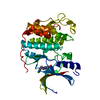
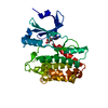
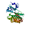
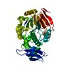
| Entry | Database: PDB / ID: 4fif | ||||||
|---|---|---|---|---|---|---|---|
| Title | Catalytic domain of human PAK4 with RPKPLVDP peptide | ||||||
 Components Components | (Serine/threonine-protein kinase PAK 4) x 2 | ||||||
 Keywords Keywords | transferase/peptide / Serine/Threonine-protein kinase PAK4 /  Kinase domain / Protein kinase / ATP binding / Phosphorylation / transferase-peptide complex Kinase domain / Protein kinase / ATP binding / Phosphorylation / transferase-peptide complex | ||||||
| Function / homology |  Function and homology information Function and homology informationdendritic spine development / cadherin binding involved in cell-cell adhesion / Activation of RAC1 / RHOV GTPase cycle / RHOJ GTPase cycle / RHOQ GTPase cycle / regulation of MAPK cascade / RHOH GTPase cycle / CDC42 GTPase cycle / RHOU GTPase cycle ...dendritic spine development / cadherin binding involved in cell-cell adhesion / Activation of RAC1 / RHOV GTPase cycle / RHOJ GTPase cycle / RHOQ GTPase cycle / regulation of MAPK cascade / RHOH GTPase cycle / CDC42 GTPase cycle / RHOU GTPase cycle / RHOG GTPase cycle / cellular response to organic cyclic compound / RAC2 GTPase cycle / RAC3 GTPase cycle / negative regulation of endothelial cell apoptotic process / cytoskeleton organization / RAC1 GTPase cycle / regulation of cell growth / adherens junction / positive regulation of angiogenesis / cell migration / non-specific serine/threonine protein kinase / protein kinase activity / intracellular signal transduction / cell cycle / phosphorylation / focal adhesion / protein serine kinase activity / protein serine/threonine kinase activity / apoptotic process / Golgi apparatus / signal transduction / ATP binding / cytosol / cytoplasmSimilarity search - Function | ||||||
| Biological species |  Homo sapiens (human) Homo sapiens (human) | ||||||
| Method | X-RAY DIFFRACTION / SYNCHROTRON / MOLECULAR REPLACEMENT / Resolution: 2.6 Å | ||||||
 Authors Authors | Ha, B.H. / Boggon, T.J. | ||||||
 Citation Citation | Journal: Proc.Natl.Acad.Sci.USA / Year: 2012 Title: Type II p21-activated kinases (PAKs) are regulated by an autoinhibitory pseudosubstrate. Authors: Ha, B.H. / Davis, M.J. / Chen, C. / Lou, H.J. / Gao, J. / Zhang, R. / Krauthammer, M. / Halaban, R. / Schlessinger, J. / Turk, B.E. / Boggon, T.J. | ||||||
| History |
|
- Structure visualization
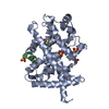
| Structure viewer | Molecule: MolmilJmol/JSmol |
|---|
- Downloads & links
-Download
| PDBx/mmCIF format | 4fif.cif.gz | 255.6 KB | Display | PDBx/mmCIF format |
|---|---|---|---|---|
| PDB format | pdb4fif.ent.gz | 206.9 KB | Display | PDB format |
| PDBx/mmJSON format | 4fif.json.gz | Tree view | PDBx/mmJSON format | |
| Others |  Other downloads Other downloads |
-Validation report
| Arichive directory | https://data.pdbj.org/pub/pdb/validation_reports/fi/4fifftp://data.pdbj.org/pub/pdb/validation_reports/fi/4fif | HTTPS FTP |
|---|
-Related structure data
| Related structure data | 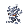 4fieC  4figC 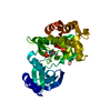 4fihC 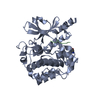 4fiiC  4fijC 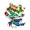 2cdzS C: citing same article ( S: Starting model for refinement |
|---|---|
| Similar structure data |


-Links
 PDBj
PDBj


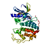
- Assembly
| Deposited unit | 
| ||||||||||||
|---|---|---|---|---|---|---|---|---|---|---|---|---|---|
| 1 | 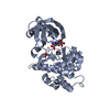
| ||||||||||||
| 2 | 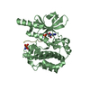
| ||||||||||||
| Unit cell |
| ||||||||||||
| Noncrystallographic symmetry (NCS) | NCS oper:
|
-Components
| #1: Protein | Mass: 39123.133 Da / Num. of mol.: 2 Source method: isolated from a genetically manipulated source Source: (gene. exp.) Homo sapiens (human) / Gene: KIAA1142, PAK4 / Plasmid: Modified pET vector / Production host:  Escherichia coli (E. coli) / Strain (production host): BL21(DE3)RILP Escherichia coli (E. coli) / Strain (production host): BL21(DE3)RILPReferences: UniProt: O96013, non-specific serine/threonine protein kinase#2: Protein/peptide | Mass: 923.110 Da / Num. of mol.: 2 / Source method: obtained synthetically / Details: Synthesized peptide sequence: RPKPLVDP / Source: (synth.) Homo sapiens (human)References: UniProt: O96013, non-specific serine/threonine protein kinase#3: Chemical |   Mass: 506.196 Da / Num. of mol.: 2 / Source method: obtained synthetically / Formula: C10H17N6O12P3 / Comment: AMP-PNP, energy-carrying molecule analogue*YM Mass: 506.196 Da / Num. of mol.: 2 / Source method: obtained synthetically / Formula: C10H17N6O12P3 / Comment: AMP-PNP, energy-carrying molecule analogue*YM#4: Water | ChemComp-HOH / | Water Mass: 18.015 Da / Num. of mol.: 56 / Source method: isolated from a natural source / Formula: H2O Mass: 18.015 Da / Num. of mol.: 56 / Source method: isolated from a natural source / Formula: H2OSequence details | SEQUENCE CORRESPONDS TO ISOFORM 2 OF SERINE/THREONINE-PROTEIN KINASE PAK 4, UNP ACCESSION CODE ...SEQUENCE CORRESPOND | |
|---|
-Experimental details
-Experiment
| Experiment | Method: X-RAY DIFFRACTION / Number of used crystals: 1 |
|---|
- Sample preparation
| Crystal | Density Matthews: 4.44 Å3/Da / Density % sol: 72.31 % |
|---|---|
| Crystal grow | Temperature: 295 K / Method: vapor diffusion, hanging drop / pH: 6 Details: 0.1M MES, 700 mM K/Na tartrate, pH 6.0, VAPOR DIFFUSION, HANGING DROP, temperature 295K |
-Data collection
| Diffraction | Mean temperature: 100 K |
|---|---|
| Diffraction source | Source: SYNCHROTRON / Site: NSLS  / Beamline: X29A / Wavelength: 1.075 Å / Beamline: X29A / Wavelength: 1.075 Å |
| Detector | Type: ADSC QUANTUM 315r / Detector: CCD / Date: Jan 30, 2012 / Details: insertion device |
| Radiation | Monochromator: Si(111) / Protocol: SINGLE WAVELENGTH / Monochromatic (M) / Laue (L): M / Scattering type: x-ray |
| Radiation wavelength | Wavelength: 1.075 Å / Relative weight: 1 |
| Reflection | Resolution: 2.6→50 Å / % possible obs: 99.6 % / Observed criterion σ(F): 0 / Observed criterion σ(I): -3 / Redundancy: 2.5 % / Biso Wilson estimate: 65.1 Å2 / Rmerge(I) obs: 0.088 / Rsym value: 0.088 / Net I/σ(I): 17.5 |
| Reflection shell | Resolution: 2.6→2.69 Å / Redundancy: 2.2 % / Rmerge(I) obs: 0.887 / Mean I/σ(I) obs: 1.8 / Rsym value: 0.887 / % possible all: 98.9 |
- Processing
| Software |
| ||||||||||||||||||||||||||||||||||||||||||||||||||||||||||||||||||||||||||||||||||||||||||||||||||||||||||||||||||||||||||||||||||||||||||||||||||||||||||||||||||||||||||
|---|---|---|---|---|---|---|---|---|---|---|---|---|---|---|---|---|---|---|---|---|---|---|---|---|---|---|---|---|---|---|---|---|---|---|---|---|---|---|---|---|---|---|---|---|---|---|---|---|---|---|---|---|---|---|---|---|---|---|---|---|---|---|---|---|---|---|---|---|---|---|---|---|---|---|---|---|---|---|---|---|---|---|---|---|---|---|---|---|---|---|---|---|---|---|---|---|---|---|---|---|---|---|---|---|---|---|---|---|---|---|---|---|---|---|---|---|---|---|---|---|---|---|---|---|---|---|---|---|---|---|---|---|---|---|---|---|---|---|---|---|---|---|---|---|---|---|---|---|---|---|---|---|---|---|---|---|---|---|---|---|---|---|---|---|---|---|---|---|---|---|---|
| Refinement | Method to determine structure: MOLECULAR REPLACEMENT Starting model: PDB ENTRY 2CDZ Resolution: 2.6→46.46 Å / Cor.coef. Fo:Fc: 0.949 / Cor.coef. Fo:Fc free: 0.938 / SU B: 19.663 / SU ML: 0.195 / Cross valid method: THROUGHOUT / ESU R: 0.268 / ESU R Free: 0.211 / Stereochemistry target values: MAXIMUM LIKELIHOOD / Details: HYDROGENS HAVE BEEN USED IF PRESENT IN THE INPUT
| ||||||||||||||||||||||||||||||||||||||||||||||||||||||||||||||||||||||||||||||||||||||||||||||||||||||||||||||||||||||||||||||||||||||||||||||||||||||||||||||||||||||||||
| Solvent computation | Ion probe radii: 0.8 Å / Shrinkage radii: 0.8 Å / VDW probe radii: 1.2 Å / Solvent model: MASK | ||||||||||||||||||||||||||||||||||||||||||||||||||||||||||||||||||||||||||||||||||||||||||||||||||||||||||||||||||||||||||||||||||||||||||||||||||||||||||||||||||||||||||
| Displacement parameters | Biso mean: 70.709 Å2
| ||||||||||||||||||||||||||||||||||||||||||||||||||||||||||||||||||||||||||||||||||||||||||||||||||||||||||||||||||||||||||||||||||||||||||||||||||||||||||||||||||||||||||
| Refinement step | Cycle: LAST / Resolution: 2.6→46.46 Å
| ||||||||||||||||||||||||||||||||||||||||||||||||||||||||||||||||||||||||||||||||||||||||||||||||||||||||||||||||||||||||||||||||||||||||||||||||||||||||||||||||||||||||||
| Refine LS restraints |
| ||||||||||||||||||||||||||||||||||||||||||||||||||||||||||||||||||||||||||||||||||||||||||||||||||||||||||||||||||||||||||||||||||||||||||||||||||||||||||||||||||||||||||
| LS refinement shell | Resolution: 2.604→2.672 Å / Total num. of bins used: 20
| ||||||||||||||||||||||||||||||||||||||||||||||||||||||||||||||||||||||||||||||||||||||||||||||||||||||||||||||||||||||||||||||||||||||||||||||||||||||||||||||||||||||||||
| Refinement TLS params. | Method: refined / Refine-ID: X-RAY DIFFRACTION
| ||||||||||||||||||||||||||||||||||||||||||||||||||||||||||||||||||||||||||||||||||||||||||||||||||||||||||||||||||||||||||||||||||||||||||||||||||||||||||||||||||||||||||
| Refinement TLS group |
|
