 Movie
Movie Controller


+ Open data

- Basic information
| Entry | Database: PDB / ID: 3mfu | ||||||
|---|---|---|---|---|---|---|---|
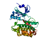
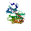
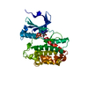
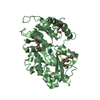
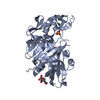
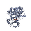
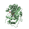
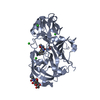
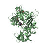
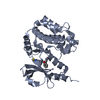
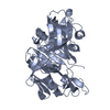
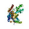
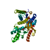
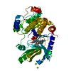
| Title | CASK-4M CaM Kinase Domain, AMPPNP-Mn2+ | ||||||
 Components Components | Peripheral plasma membrane protein CASK | ||||||
 Keywords Keywords |  TRANSFERASE / Catalytic mechanism / kinase catalysis / Mg2+-mediated phosphate transfer / protein kinase TRANSFERASE / Catalytic mechanism / kinase catalysis / Mg2+-mediated phosphate transfer / protein kinase | ||||||
| Function / homology |  Function and homology information Function and homology informationnegative regulation of cellular response to growth factor stimulus / guanylate kinase activity / Dopamine Neurotransmitter Release Cycle / neurexin family protein binding / regulation of neurotransmitter secretion / negative regulation of wound healing / nuclear lamina / calcium ion import / Assembly and cell surface presentation of NMDA receptors / Neurexins and neuroligins ...negative regulation of cellular response to growth factor stimulus / guanylate kinase activity / Dopamine Neurotransmitter Release Cycle / neurexin family protein binding / regulation of neurotransmitter secretion / negative regulation of wound healing / nuclear lamina / calcium ion import / Assembly and cell surface presentation of NMDA receptors / Neurexins and neuroligins / Sensory processing of sound by outer hair cells of the cochlea / Sensory processing of sound by inner hair cells of the cochlea / Nephrin family interactions / ciliary membrane / regulation of synaptic vesicle exocytosis / Syndecan interactions / negative regulation of cell-matrix adhesion / positive regulation of calcium ion import / basement membrane / negative regulation of keratinocyte proliferation / establishment of localization in cell / Schaffer collateral - CA1 synapse / nuclear matrix / cell-cell junction / actin cytoskeleton / presynaptic membrane / basolateral plasma membrane / vesicle / calmodulin binding / non-specific serine/threonine protein kinase / cell adhesion / phosphorylation / signaling receptor binding / focal adhesion / protein serine kinase activity / protein serine/threonine kinase activity / nucleolus / positive regulation of transcription by RNA polymerase II / ATP binding / plasma membrane / cytosol / cytoplasmSimilarity search - Function | ||||||
| Biological species |  Homo sapiens (human) Homo sapiens (human) | ||||||
| Method | X-RAY DIFFRACTION / SYNCHROTRON / MOLECULAR REPLACEMENT / Resolution: 2.3 Å | ||||||
 Authors Authors | Wahl, M.C. / Mukherjee, K. | ||||||
 Citation Citation | Journal: Sci.Signal. / Year: 2010 Title: Evolution of CASK into a Mg2+-sensitive kinase. Authors: Mukherjee, K. / Sharma, M. / Jahn, R. / Wahl, M.C. / Sudhof, T.C. | ||||||
| History |
|
- Structure visualization
| Structure viewer | Molecule: MolmilJmol/JSmol |
|---|
- Downloads & links
-Download
| PDBx/mmCIF format | 3mfu.cif.gz | 82.6 KB | Display | PDBx/mmCIF format |
|---|---|---|---|---|
| PDB format | pdb3mfu.ent.gz | 58.9 KB | Display | PDB format |
| PDBx/mmJSON format | 3mfu.json.gz | Tree view | PDBx/mmJSON format | |
| Others |  Other downloads Other downloads |
-Validation report
| Arichive directory | https://data.pdbj.org/pub/pdb/validation_reports/mf/3mfuftp://data.pdbj.org/pub/pdb/validation_reports/mf/3mfu | HTTPS FTP |
|---|
-Related structure data
| Related structure data |  3mfrC  3mfsC  3mftC 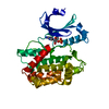 3c0iS S: Starting model for refinement C: citing same article ( |
|---|---|
| Similar structure data |


-Links
 PDBj
PDBj


- Assembly
| Deposited unit | 
| ||||||||
|---|---|---|---|---|---|---|---|---|---|
| 1 |
| ||||||||
| Unit cell |
|
-Components
| #1: Protein | Mass: 39291.047 Da / Num. of mol.: 1 / Fragment: CASK-4M CaM kinase domain, residues 1-337 / Mutation: Pro22Ala, His145Glu, Gly162Asp, Cys146Asn Source method: isolated from a genetically manipulated source Source: (gene. exp.) Homo sapiens (human) / Gene: CASK, LIN2 / Plasmid: pGEX / Production host:  Escherichia coli (E. coli) / Strain (production host): BL21(DE3) Escherichia coli (E. coli) / Strain (production host): BL21(DE3)References: UniProt: O14936, non-specific serine/threonine protein kinase |
|---|---|
| #2: Chemical | ChemComp-ANP /   Mass: 506.196 Da / Num. of mol.: 1 / Source method: obtained synthetically / Formula: C10H17N6O12P3 / Comment: AMP-PNP, energy-carrying molecule analogue*YM Mass: 506.196 Da / Num. of mol.: 1 / Source method: obtained synthetically / Formula: C10H17N6O12P3 / Comment: AMP-PNP, energy-carrying molecule analogue*YM |
| #3: Chemical | ChemComp-MN /   Mass: 54.938 Da / Num. of mol.: 1 / Source method: obtained synthetically / Formula: Mn Mass: 54.938 Da / Num. of mol.: 1 / Source method: obtained synthetically / Formula: Mn |
| #4: Water | ChemComp-HOH / Water Mass: 18.015 Da / Num. of mol.: 190 / Source method: isolated from a natural source / Formula: H2O Mass: 18.015 Da / Num. of mol.: 190 / Source method: isolated from a natural source / Formula: H2O |
-Experimental details
-Experiment
| Experiment | Method: X-RAY DIFFRACTION / Number of used crystals: 1 |
|---|
- Sample preparation
| Crystal | Density Matthews: 2.34 Å3/Da / Density % sol: 47.39 % |
|---|---|
| Crystal grow | Temperature: 293 K / Method: vapor diffusion, sitting drop / pH: 7.2 Details: 12.5 % (v/v) ethylene glycol, pH 7.2, VAPOR DIFFUSION, SITTING DROP, temperature 293K |
-Data collection
| Diffraction | Mean temperature: 100 K |
|---|---|
| Diffraction source | Source: SYNCHROTRON / Site: BESSY  / Beamline: 14.2 / Wavelength: 1.87856 Å / Beamline: 14.2 / Wavelength: 1.87856 Å |
| Detector | Type: MARMOSAIC 225 mm CCD / Detector: CCD |
| Radiation | Monochromator: Si 111 CHANNEL / Protocol: SINGLE WAVELENGTH / Monochromatic (M) / Laue (L): M / Scattering type: x-ray |
| Radiation wavelength | Wavelength: 1.87856 Å / Relative weight: 1 |
| Reflection | Resolution: 2.3→20 Å / Num. all: 31627 / % possible obs: 98.2 % / Observed criterion σ(F): 1 / Observed criterion σ(I): 1 / Redundancy: 4.5 % / Biso Wilson estimate: 51.1 Å2 / Rmerge(I) obs: 0.063 / Rsym value: 0.063 / Net I/σ(I): 14.4 |
| Reflection shell | Resolution: 2.3→2.36 Å / Redundancy: 2.7 % / Rmerge(I) obs: 0.497 / Mean I/σ(I) obs: 2.2 / Num. unique all: 1968 / Rsym value: 0.497 / % possible all: 83.8 |
- Processing
| Software |
| ||||||||||||||||||||||||||||||||||||||||||||||||||||||||||||||||||||||||||||||||||||||||||
|---|---|---|---|---|---|---|---|---|---|---|---|---|---|---|---|---|---|---|---|---|---|---|---|---|---|---|---|---|---|---|---|---|---|---|---|---|---|---|---|---|---|---|---|---|---|---|---|---|---|---|---|---|---|---|---|---|---|---|---|---|---|---|---|---|---|---|---|---|---|---|---|---|---|---|---|---|---|---|---|---|---|---|---|---|---|---|---|---|---|---|---|
| Refinement | Method to determine structure: MOLECULAR REPLACEMENT Starting model: 3C0I Resolution: 2.3→19.65 Å / Cor.coef. Fo:Fc: 0.957 / Cor.coef. Fo:Fc free: 0.934 / SU B: 13.302 / SU ML: 0.174 / TLS residual ADP flag: LIKELY RESIDUAL / Cross valid method: THROUGHOUT / σ(F): 0 / σ(I): 0 / ESU R: 0.298 / ESU R Free: 0.224 / Stereochemistry target values: MAXIMUM LIKELIHOOD / Details: HYDROGENS HAVE BEEN ADDED IN THE RIDING POSITIONS
| ||||||||||||||||||||||||||||||||||||||||||||||||||||||||||||||||||||||||||||||||||||||||||
| Solvent computation | Ion probe radii: 0.8 Å / Shrinkage radii: 0.8 Å / VDW probe radii: 1.4 Å / Solvent model: MASK | ||||||||||||||||||||||||||||||||||||||||||||||||||||||||||||||||||||||||||||||||||||||||||
| Displacement parameters | Biso mean: 43.539 Å2
| ||||||||||||||||||||||||||||||||||||||||||||||||||||||||||||||||||||||||||||||||||||||||||
| Refine analyze | Luzzati coordinate error obs: 0.17 Å | ||||||||||||||||||||||||||||||||||||||||||||||||||||||||||||||||||||||||||||||||||||||||||
| Refinement step | Cycle: LAST / Resolution: 2.3→19.65 Å
| ||||||||||||||||||||||||||||||||||||||||||||||||||||||||||||||||||||||||||||||||||||||||||
| Refine LS restraints |
| ||||||||||||||||||||||||||||||||||||||||||||||||||||||||||||||||||||||||||||||||||||||||||
| LS refinement shell | Resolution: 2.3→2.359 Å / Total num. of bins used: 20
| ||||||||||||||||||||||||||||||||||||||||||||||||||||||||||||||||||||||||||||||||||||||||||
| Refinement TLS params. | Method: refined / Refine-ID: X-RAY DIFFRACTION
| ||||||||||||||||||||||||||||||||||||||||||||||||||||||||||||||||||||||||||||||||||||||||||
| Refinement TLS group |
|
