 Movie
Movie Controller

[English] 日本語
 Yorodumi
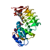
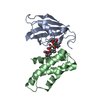
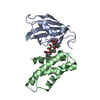
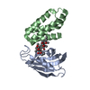
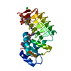
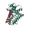
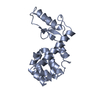
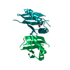
- PDB-4fap: ATOMIC STRUCTURES OF THE RAPAMYCIN ANALOGS IN COMPLEX WITH BOTH H... -
+ Open data

- Basic information
| Entry | Database: PDB / ID: 4fap | ||||||
|---|---|---|---|---|---|---|---|
| Title | ATOMIC STRUCTURES OF THE RAPAMYCIN ANALOGS IN COMPLEX WITH BOTH HUMAN FKBP12 AND FRB DOMAIN OF FRAP | ||||||
 Components Components |
| ||||||
 Keywords Keywords |  CELL CYCLE / FKBP12 / FRAP / RAPAMYCIN / COMPLEX / GENE THERAPY CELL CYCLE / FKBP12 / FRAP / RAPAMYCIN / COMPLEX / GENE THERAPY | ||||||
| Function / homology |  Function and homology information Function and homology informationRNA polymerase III type 2 promoter sequence-specific DNA binding / positive regulation of cytoplasmic translational initiation / RNA polymerase III type 1 promoter sequence-specific DNA binding / positive regulation of pentose-phosphate shunt / T-helper 1 cell lineage commitment / regulation of locomotor rhythm / positive regulation of wound healing, spreading of epidermal cells / cellular response to leucine starvation / TFIIIC-class transcription factor complex binding / TORC2 complex ...RNA polymerase III type 2 promoter sequence-specific DNA binding / positive regulation of cytoplasmic translational initiation / RNA polymerase III type 1 promoter sequence-specific DNA binding / positive regulation of pentose-phosphate shunt / T-helper 1 cell lineage commitment / regulation of locomotor rhythm / positive regulation of wound healing, spreading of epidermal cells / cellular response to leucine starvation / TFIIIC-class transcription factor complex binding / TORC2 complex / regulation of membrane permeability / heart valve morphogenesis / negative regulation of lysosome organization / macrolide binding / RNA polymerase III type 3 promoter sequence-specific DNA binding / TORC1 complex / positive regulation of transcription of nucleolar large rRNA by RNA polymerase I / calcineurin-NFAT signaling cascade / activin receptor binding / cytoplasmic side of membrane / regulation of autophagosome assembly / TORC1 signaling / voluntary musculoskeletal movement / regulation of osteoclast differentiation / positive regulation of keratinocyte migration / transforming growth factor beta receptor binding / TGFBR1 LBD Mutants in Cancer / cellular response to L-leucine / signaling receptor inhibitor activity / MTOR signalling / Amino acids regulate mTORC1 / cellular response to nutrient / type I transforming growth factor beta receptor binding / energy reserve metabolic process / Energy dependent regulation of mTOR by LKB1-AMPK / nucleus localization / negative regulation of activin receptor signaling pathway / ruffle organization / heart trabecula formation / negative regulation of cell size / cellular response to osmotic stress / terminal cisterna / ryanodine receptor complex / I-SMAD binding / regulation of amyloid precursor protein catabolic process / anoikis / cardiac muscle cell development / negative regulation of protein localization to nucleus / positive regulation of transcription by RNA polymerase III / protein maturation by protein folding / regulation of myelination / negative regulation of calcineurin-NFAT signaling cascade / 'de novo' protein folding / ventricular cardiac muscle tissue morphogenesis / Macroautophagy / regulation of cell size / negative regulation of macroautophagy / lysosome organization / negative regulation of phosphoprotein phosphatase activity / positive regulation of oligodendrocyte differentiation / FK506 binding / positive regulation of actin filament polymerization / positive regulation of myotube differentiation / behavioral response to pain / TOR signaling / oligodendrocyte differentiation / mTORC1-mediated signalling / TGF-beta receptor signaling activates SMADs / Constitutive Signaling by AKT1 E17K in Cancer / germ cell development / cellular response to nutrient levels / CD28 dependent PI3K/Akt signaling / positive regulation of translational initiation / positive regulation of phosphoprotein phosphatase activity / neuronal action potential / Calcineurin activates NFAT / HSF1-dependent transactivation / regulation of macroautophagy / positive regulation of epithelial to mesenchymal transition / endomembrane system / regulation of immune response / 'de novo' pyrimidine nucleobase biosynthetic process / response to amino acid / protein peptidyl-prolyl isomerization / supramolecular fiber organization / positive regulation of lamellipodium assembly / phagocytic vesicle / positive regulation of lipid biosynthetic process / regulation of cellular response to heat / heart morphogenesis / regulation of ryanodine-sensitive calcium-release channel activity / cardiac muscle contraction / positive regulation of stress fiber assembly / cytoskeleton organization / sarcoplasmic reticulum membrane / cellular response to amino acid starvation / T cell costimulation / cellular response to starvation / positive regulation of glycolytic process / T cell activationSimilarity search - Function | ||||||
| Biological species |  Homo sapiens (human) Homo sapiens (human) | ||||||
| Method | X-RAY DIFFRACTION / MOLECULAR REPLACEMENT / Resolution: 2.8 Å | ||||||
 Authors Authors | Liang, J. / Clardy, J. | ||||||
 Citation Citation | Journal: Acta Crystallogr.,Sect.D / Year: 1999 Title: Refined structure of the FKBP12-rapamycin-FRB ternary complex at 2.2 A resolution. Authors: Liang, J. / Choi, J. / Clardy, J. | ||||||
| History |
|
- Structure visualization
| Structure viewer | Molecule: MolmilJmol/JSmol |
|---|
- Downloads & links
-Download
| PDBx/mmCIF format | 4fap.cif.gz | 56 KB | Display | PDBx/mmCIF format |
|---|---|---|---|---|
| PDB format | pdb4fap.ent.gz | 39.3 KB | Display | PDB format |
| PDBx/mmJSON format | 4fap.json.gz | Tree view | PDBx/mmJSON format | |
| Others |  Other downloads Other downloads |
-Validation report
| Arichive directory | https://data.pdbj.org/pub/pdb/validation_reports/fa/4fapftp://data.pdbj.org/pub/pdb/validation_reports/fa/4fap | HTTPS FTP |
|---|
-Related structure data
| Related structure data |  1nsgC  2fapSC  3fapC S: Starting model for refinement C: citing same article ( |
|---|---|
| Similar structure data |


-Links
 PDBj
PDBj


- Assembly
| Deposited unit | 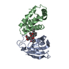
| ||||||||
|---|---|---|---|---|---|---|---|---|---|
| 1 |
| ||||||||
| Unit cell |
|
-Components
| #1: Protein | Mass: 11836.508 Da / Num. of mol.: 1 Source method: isolated from a genetically manipulated source Source: (gene. exp.) Homo sapiens (human) / Strain: BL21 (DE3) (NOVAGEN)Gene: HUMAN HIPPOCAMPAL CDNA LIBRARY SOURCE 8 (CLONTECH, PALO ALTO, CA) Plasmid: PGEX-3X / Production host:  Escherichia coli (E. coli) / Strain (production host): BL21 (DE3) (NOVAGEN) / References: UniProt: P62942, peptidylprolyl isomerase Escherichia coli (E. coli) / Strain (production host): BL21 (DE3) (NOVAGEN) / References: UniProt: P62942, peptidylprolyl isomerase |
|---|---|
| #2: Protein | Mass: 11331.937 Da / Num. of mol.: 1 / Fragment: FRB / Source method: isolated from a natural source / Source: (natural) Homo sapiens (human) / References: UniProt: P42345 |
| #3: Chemical | ChemComp-ARD /   Mass: 980.296 Da / Num. of mol.: 1 / Source method: obtained synthetically / Formula: C55H81NO12S / Comment: antibiotic*YM Mass: 980.296 Da / Num. of mol.: 1 / Source method: obtained synthetically / Formula: C55H81NO12S / Comment: antibiotic*YM |
-Experimental details
-Experiment
| Experiment | Method: X-RAY DIFFRACTION / Number of used crystals: 1 |
|---|
- Sample preparation
| Crystal | Density Matthews: 2.62 Å3/Da / Density % sol: 50 % |
|---|---|
| Crystal grow | pH: 8 Details: 20% PEG8000, 10% MPD, 0.1 M TRIS-HCL PH 8.5, pH 8.0 |
| Crystal grow | *PLUS Method: otherDetails: This particular structure is not described in this paper. |
-Data collection
| Diffraction | Mean temperature: 298 K |
|---|---|
| Diffraction source | Source: ROTATING ANODE / Type: RIGAKU RU200 / Wavelength: 1.5418 |
| Detector | Type: SDMS / Detector: AREA DETECTOR / Date: Feb 1, 1997 |
| Radiation | Monochromator: NI FILTER / Protocol: SINGLE WAVELENGTH / Monochromatic (M) / Laue (L): M / Scattering type: x-ray |
| Radiation wavelength | Wavelength: 1.5418 Å / Relative weight: 1 |
| Reflection | Resolution: 2.8→20 Å / Num. obs: 5598 / % possible obs: 91 % / Observed criterion σ(I): 0 / Redundancy: 3.1 % / Rsym value: 8.6 / Net I/σ(I): 10.4 |
- Processing
| Software |
| ||||||||||||||||||||||||||||||||||||||||||||||||||||||||||||||||||||||||||||||||
|---|---|---|---|---|---|---|---|---|---|---|---|---|---|---|---|---|---|---|---|---|---|---|---|---|---|---|---|---|---|---|---|---|---|---|---|---|---|---|---|---|---|---|---|---|---|---|---|---|---|---|---|---|---|---|---|---|---|---|---|---|---|---|---|---|---|---|---|---|---|---|---|---|---|---|---|---|---|---|---|---|---|
| Refinement | Method to determine structure: MOLECULAR REPLACEMENT Starting model: 2FAP Resolution: 2.8→20 Å / Rfactor Rfree error: 0.012 / Data cutoff high rms absF: 72314.66 / Isotropic thermal model: RESTRAINED / Cross valid method: THROUGHOUT / σ(F): 0
| ||||||||||||||||||||||||||||||||||||||||||||||||||||||||||||||||||||||||||||||||
| Solvent computation | Solvent model: FLAT MODEL / Bsol: 11.96 Å2 / ksol: 0.271 e/Å3 | ||||||||||||||||||||||||||||||||||||||||||||||||||||||||||||||||||||||||||||||||
| Displacement parameters | Biso mean: 32.6 Å2
| ||||||||||||||||||||||||||||||||||||||||||||||||||||||||||||||||||||||||||||||||
| Refine analyze |
| ||||||||||||||||||||||||||||||||||||||||||||||||||||||||||||||||||||||||||||||||
| Refinement step | Cycle: LAST / Resolution: 2.8→20 Å
| ||||||||||||||||||||||||||||||||||||||||||||||||||||||||||||||||||||||||||||||||
| Refine LS restraints |
| ||||||||||||||||||||||||||||||||||||||||||||||||||||||||||||||||||||||||||||||||
| LS refinement shell | Resolution: 2.8→2.97 Å / Rfactor Rfree error: 0.018 / Total num. of bins used: 6
| ||||||||||||||||||||||||||||||||||||||||||||||||||||||||||||||||||||||||||||||||
| Xplor file |
|
