 Movie
Movie Controller

[English] 日本語
 Yorodumi
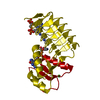
- PDB-7l7z: x-ray structure of the N-acetyltransferase Pcryo_0637 from psychr... -
+ Open data

- Basic information
| Entry | Database: PDB / ID: 7l7z | ||||||
|---|---|---|---|---|---|---|---|
| Title | x-ray structure of the N-acetyltransferase Pcryo_0637 from psychrobacter cryohalolentis in the presence of coenzyme A and UDP-di-N-acetyl-bacillosamine | ||||||
 Components Components | Putative acetyl transferase protein | ||||||
 Keywords Keywords |  TRANSFERASE / beta helix / bacillosamine / lipopolysaccharide / carbohydrate TRANSFERASE / beta helix / bacillosamine / lipopolysaccharide / carbohydrate | ||||||
| Function / homology | Sialic acid O-acyltransferase NeuD-like / PglD, N-terminal / PglD N-terminal domain / Trimeric LpxA-like superfamily / transferase activity / COENZYME A / UDP-di-N-acetyl-alpha-bacillosamine / Acetyl transferase protein Function and homology information Function and homology information | ||||||
| Biological species |  Psychrobacter cryohalolentis (bacteria) Psychrobacter cryohalolentis (bacteria) | ||||||
| Method | X-RAY DIFFRACTION / FOURIER SYNTHESIS / Resolution: 1.55 Å | ||||||
 Authors Authors | Linehan, M.P. / Thoden, J.B. / Holden, H.M. | ||||||
| Funding support |  United States, 1items United States, 1items
| ||||||
 Citation Citation | Journal: J.Biol.Chem. / Year: 2021 Title: Characterization of two enzymes from Psychrobacter cryohalolentis that are required for the biosynthesis of an unusual diacetamido-d-sugar. Authors: Linehan, M.P. / Thoden, J.B. / Holden, H.M. | ||||||
| History |
|
- Structure visualization
| Structure viewer | Molecule: MolmilJmol/JSmol |
|---|
- Downloads & links
-Download
| PDBx/mmCIF format | 7l7z.cif.gz | 70.4 KB | Display | PDBx/mmCIF format |
|---|---|---|---|---|
| PDB format | pdb7l7z.ent.gz | Display | PDB format | |
| PDBx/mmJSON format | 7l7z.json.gz | Tree view | PDBx/mmJSON format | |
| Others |  Other downloads Other downloads |
-Validation report
| Arichive directory | https://data.pdbj.org/pub/pdb/validation_reports/l7/7l7zftp://data.pdbj.org/pub/pdb/validation_reports/l7/7l7z | HTTPS FTP |
|---|
-Related structure data
| Related structure data |  7l7xC  7l7ySC  7l81C  7l82C S: Starting model for refinement C: citing same article ( |
|---|---|
| Similar structure data |


-Links
 PDBj
PDBj


- Assembly
| Deposited unit | 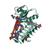
| ||||||||||||
|---|---|---|---|---|---|---|---|---|---|---|---|---|---|
| 1 | 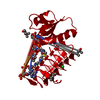
| ||||||||||||
| Unit cell |
| ||||||||||||
| Components on special symmetry positions |
|
-Components
-Protein , 1 types, 1 molecules AAA
| #1: Protein | Mass: 23680.221 Da / Num. of mol.: 1 Source method: isolated from a genetically manipulated source Source: (gene. exp.) Psychrobacter cryohalolentis (strain ATCC BAA-1226 / DSM 17306 / VKM B-2378 / K5) (bacteria)Strain: ATCC BAA-1226 / DSM 17306 / VKM B-2378 / K5 / Gene: Pcryo_0637 / Production host: Escherichia coli (E. coli) / References: UniProt: Q1QD33 |
|---|
-Non-polymers , 5 types, 233 molecules 


| #2: Chemical | ChemComp-COA / Coenzyme A Mass: 767.534 Da / Num. of mol.: 1 / Source method: obtained synthetically / Formula: C21H36N7O16P3S / Feature type: SUBJECT OF INVESTIGATION Mass: 767.534 Da / Num. of mol.: 1 / Source method: obtained synthetically / Formula: C21H36N7O16P3S / Feature type: SUBJECT OF INVESTIGATION | ||
|---|---|---|---|
| #3: Chemical | ChemComp-XQA /  Mass: 632.406 Da / Num. of mol.: 1 / Source method: obtained synthetically / Formula: C19H30N4O16P2 / Feature type: SUBJECT OF INVESTIGATION Mass: 632.406 Da / Num. of mol.: 1 / Source method: obtained synthetically / Formula: C19H30N4O16P2 / Feature type: SUBJECT OF INVESTIGATION | ||
| #4: Chemical | ChemComp-CL / Chloride Mass: 35.453 Da / Num. of mol.: 1 / Source method: obtained synthetically / Formula: Cl Mass: 35.453 Da / Num. of mol.: 1 / Source method: obtained synthetically / Formula: Cl | ||
| #5: Chemical | ChemComp-MPD / ( 2-Methyl-2,4-pentanediol Mass: 118.174 Da / Num. of mol.: 5 / Source method: obtained synthetically / Formula: C6H14O2 / Comment: precipitant*YM Mass: 118.174 Da / Num. of mol.: 5 / Source method: obtained synthetically / Formula: C6H14O2 / Comment: precipitant*YM#6: Water | ChemComp-HOH / | WaterMass: 18.015 Da / Num. of mol.: 225 / Source method: isolated from a natural source / Formula: H2O |
-Details
| Has ligand of interest | Y |
|---|
-Experimental details
-Experiment
| Experiment | Method: X-RAY DIFFRACTION / Number of used crystals: 1 |
|---|
- Sample preparation
| Crystal | Density Matthews: 2.54 Å3/Da / Density % sol: 51.62 % |
|---|---|
| Crystal grow | Temperature: 293 K / Method: vapor diffusion, hanging drop / pH: 7.5 Details: 23-26% MPD, 100 mM HEPES. protein incubated with 5 mM coenzyme A and 20 mM UDP-2,4-diacetamido-2,4,6-trideoxy-D-glucose |
-Data collection
| Diffraction | Mean temperature: 100 K / Serial crystal experiment: N |
|---|---|
| Diffraction source | Source: SEALED TUBE / Type: BRUKER D8 QUEST / Wavelength: 1.54718 Å |
| Detector | Type: Bruker PHOTON II / Detector: PIXEL / Date: Sep 19, 2019 |
| Radiation | Protocol: SINGLE WAVELENGTH / Monochromatic (M) / Laue (L): M / Scattering type: x-ray |
| Radiation wavelength | Wavelength: 1.54718 Å / Relative weight: 1 |
| Reflection | Resolution: 1.55→50 Å / Num. obs: 33312 / % possible obs: 98.1 % / Observed criterion σ(F): 0 / Observed criterion σ(I): 0 / Redundancy: 5.1 % / Rsym value: 0.045 / Net I/σ(I): 19.6 |
| Reflection shell | Resolution: 1.55→1.65 Å / Redundancy: 2.4 % / Mean I/σ(I) obs: 3 / Num. unique obs: 5566 / Rsym value: 0.341 / % possible all: 95 |
- Processing
| Software |
| |||||||||||||||||||||||||||||||||||||||||||||||||||||||||||||||||||||||||||||||||||||||||||||||||||||||||||||||||||||||||||||||||||||||||||||||||||||||||||
|---|---|---|---|---|---|---|---|---|---|---|---|---|---|---|---|---|---|---|---|---|---|---|---|---|---|---|---|---|---|---|---|---|---|---|---|---|---|---|---|---|---|---|---|---|---|---|---|---|---|---|---|---|---|---|---|---|---|---|---|---|---|---|---|---|---|---|---|---|---|---|---|---|---|---|---|---|---|---|---|---|---|---|---|---|---|---|---|---|---|---|---|---|---|---|---|---|---|---|---|---|---|---|---|---|---|---|---|---|---|---|---|---|---|---|---|---|---|---|---|---|---|---|---|---|---|---|---|---|---|---|---|---|---|---|---|---|---|---|---|---|---|---|---|---|---|---|---|---|---|---|---|---|---|---|---|---|
| Refinement | Method to determine structure: FOURIER SYNTHESIS Starting model: 7l7y Resolution: 1.55→30.983 Å / Cor.coef. Fo:Fc: 0.973 / Cor.coef. Fo:Fc free: 0.965 / SU B: 1.614 / SU ML: 0.055 / Cross valid method: FREE R-VALUE / ESU R: 0.075 / ESU R Free: 0.076 Details: Hydrogens have been added in their riding positions
| |||||||||||||||||||||||||||||||||||||||||||||||||||||||||||||||||||||||||||||||||||||||||||||||||||||||||||||||||||||||||||||||||||||||||||||||||||||||||||
| Solvent computation | Ion probe radii: 0.8 Å / Shrinkage radii: 0.8 Å / VDW probe radii: 1.2 Å / Solvent model: MASK BULK SOLVENT | |||||||||||||||||||||||||||||||||||||||||||||||||||||||||||||||||||||||||||||||||||||||||||||||||||||||||||||||||||||||||||||||||||||||||||||||||||||||||||
| Displacement parameters | Biso mean: 18.657 Å2
| |||||||||||||||||||||||||||||||||||||||||||||||||||||||||||||||||||||||||||||||||||||||||||||||||||||||||||||||||||||||||||||||||||||||||||||||||||||||||||
| Refinement step | Cycle: LAST / Resolution: 1.55→30.983 Å
| |||||||||||||||||||||||||||||||||||||||||||||||||||||||||||||||||||||||||||||||||||||||||||||||||||||||||||||||||||||||||||||||||||||||||||||||||||||||||||
| Refine LS restraints |
| |||||||||||||||||||||||||||||||||||||||||||||||||||||||||||||||||||||||||||||||||||||||||||||||||||||||||||||||||||||||||||||||||||||||||||||||||||||||||||
| LS refinement shell |
|
