 Movie
Movie Controller

[English] 日本語
 Yorodumi
- PDB-4ea8: X-ray crystal structure of PerB from Caulobacter crescentus in co... -
+ Open data

- Basic information
| Entry | Database: PDB / ID: 4ea8 | ||||||
|---|---|---|---|---|---|---|---|
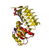
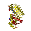
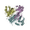
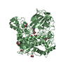
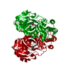
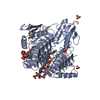
| Title | X-ray crystal structure of PerB from Caulobacter crescentus in complex with coenzyme A and GDP-N-acetylperosamine at 1 Angstrom resolution | ||||||
 Components Components | Perosamine N-acetyltransferase | ||||||
 Keywords Keywords |  TRANSFERASE / beta helix / acetyltransferase / acetyl coenzyme A / GDP-perosamine TRANSFERASE / beta helix / acetyltransferase / acetyl coenzyme A / GDP-perosamine | ||||||
| Function / homology |  Function and homology information Function and homology information | ||||||
| Biological species |  Caulobacter vibrioides (bacteria) Caulobacter vibrioides (bacteria) | ||||||
| Method | X-RAY DIFFRACTION / SYNCHROTRON / MOLECULAR REPLACEMENT / Resolution: 1 Å | ||||||
 Authors Authors | Thoden, J.B. / Reinhardt, L.A. / Cook, P.D. / Menden, P. / Cleland, W.W. / Holden, H.M. | ||||||
 Citation Citation | Journal: Biochemistry / Year: 2012 Title: Catalytic Mechanism of Perosamine N-Acetyltransferase Revealed by High-Resolution X-ray Crystallographic Studies and Kinetic Analyses. Authors: Thoden, J.B. / Reinhardt, L.A. / Cook, P.D. / Menden, P. / Cleland, W.W. / Holden, H.M. | ||||||
| History |
|
- Structure visualization
| Structure viewer | Molecule: MolmilJmol/JSmol |
|---|
- Downloads & links
-Download
| PDBx/mmCIF format | 4ea8.cif.gz | 111.7 KB | Display | PDBx/mmCIF format |
|---|---|---|---|---|
| PDB format | pdb4ea8.ent.gz | 84 KB | Display | PDB format |
| PDBx/mmJSON format | 4ea8.json.gz | Tree view | PDBx/mmJSON format | |
| Others |  Other downloads Other downloads |
-Validation report
| Arichive directory | https://data.pdbj.org/pub/pdb/validation_reports/ea/4ea8ftp://data.pdbj.org/pub/pdb/validation_reports/ea/4ea8 | HTTPS FTP |
|---|
-Related structure data
| Related structure data |  4ea7SC  4ea9C  4eaaC  4eabC S: Starting model for refinement C: citing same article ( |
|---|---|
| Similar structure data |


-Links
 PDBj
PDBj


- Assembly
| Deposited unit | 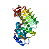
| ||||||||
|---|---|---|---|---|---|---|---|---|---|
| 1 | 
| ||||||||
| Unit cell |
| ||||||||
| Components on special symmetry positions |
|
-Components
| #1: Protein | Mass: 21910.334 Da / Num. of mol.: 1 Source method: isolated from a genetically manipulated source Source: (gene. exp.) Caulobacter vibrioides (bacteria) / Gene: CC_1011, wbqR / Plasmid: pET28 / Production host: Escherichia coli (E. coli) / Strain (production host): Rosetta2(DE3)References: UniProt: O85353, Transferases; Acyltransferases; Transferring groups other than aminoacyl groups | ||
|---|---|---|---|
| #2: Chemical | ChemComp-COA / Coenzyme A  Mass: 767.534 Da / Num. of mol.: 1 / Source method: obtained synthetically / Formula: C21H36N7O16P3S Mass: 767.534 Da / Num. of mol.: 1 / Source method: obtained synthetically / Formula: C21H36N7O16P3S | ||
| #3: Chemical | ChemComp-JB3 /   Mass: 630.394 Da / Num. of mol.: 1 / Source method: obtained synthetically / Formula: C18H28N6O15P2 Mass: 630.394 Da / Num. of mol.: 1 / Source method: obtained synthetically / Formula: C18H28N6O15P2 | ||
| #4: Chemical | Chloride  Mass: 35.453 Da / Num. of mol.: 2 / Source method: obtained synthetically / Formula: Cl Mass: 35.453 Da / Num. of mol.: 2 / Source method: obtained synthetically / Formula: Cl#5: Water | ChemComp-HOH / | Water Mass: 18.015 Da / Num. of mol.: 334 / Source method: isolated from a natural source / Formula: H2O Mass: 18.015 Da / Num. of mol.: 334 / Source method: isolated from a natural source / Formula: H2O |
-Experimental details
-Experiment
| Experiment | Method: X-RAY DIFFRACTION / Number of used crystals: 1 |
|---|
- Sample preparation
| Crystal | Density Matthews: 2.93 Å3/Da / Density % sol: 57.98 % |
|---|---|
| Crystal grow | Temperature: 295 K / Method: vapor diffusion, hanging drop / pH: 7.5 Details: 25-30% PEG5000 MME, 100 mM HEPES, pH 7.5, 5 mM CoA, 5 mM GDP, VAPOR DIFFUSION, HANGING DROP, temperature 295K |
-Data collection
| Diffraction | Mean temperature: 100 K |
|---|---|
| Diffraction source | Source: SYNCHROTRON / Site: APS  / Beamline: 19-ID / Wavelength: 0.667 Å / Beamline: 19-ID / Wavelength: 0.667 Å |
| Detector | Type: ADSC QUANTUM 315r / Detector: CCD / Date: Nov 19, 2009 |
| Radiation | Monochromator: double crystal Si(111) / Protocol: SINGLE WAVELENGTH / Monochromatic (M) / Laue (L): M / Scattering type: x-ray |
| Radiation wavelength | Wavelength: 0.667 Å / Relative weight: 1 |
| Reflection | Resolution: 1→50 Å / Num. all: 132601 / Num. obs: 132601 / % possible obs: 98.5 % / Observed criterion σ(F): 0 / Observed criterion σ(I): 0 / Redundancy: 7.2 % / Rmerge(I) obs: 0.063 / Rsym value: 0.063 / Net I/σ(I): 47.1 |
| Reflection shell | Resolution: 1→1.02 Å / Redundancy: 3.3 % / Rmerge(I) obs: 0.391 / Mean I/σ(I) obs: 2 / Num. unique all: 6425 / Rsym value: 0.391 / % possible all: 95.8 |
- Processing
| Software |
| |||||||||||||||||||||||||
|---|---|---|---|---|---|---|---|---|---|---|---|---|---|---|---|---|---|---|---|---|---|---|---|---|---|---|
| Refinement | Method to determine structure: MOLECULAR REPLACEMENT Starting model: PDB ENTRY 4EA7 Resolution: 1→50 Å / Cross valid method: THROUGHOUT / σ(F): 0 / σ(I): 0 / Stereochemistry target values: Engh & Huber
| |||||||||||||||||||||||||
| Refinement step | Cycle: LAST / Resolution: 1→50 Å
|
