 Movie
Movie Controller


+ Open data

- Basic information
| Entry | Database: PDB / ID: 4f87 | ||||||
|---|---|---|---|---|---|---|---|
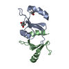
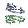
| Title | X-ray Crystal Structure of PlyCB | ||||||
 Components Components | PlyCB | ||||||
 Keywords Keywords |  ANTIMICROBIAL PROTEIN / VIRAL PROTEIN / Lysin / bacteriophage ANTIMICROBIAL PROTEIN / VIRAL PROTEIN / Lysin / bacteriophage | ||||||
| Function / homology | Signal recognition particle alu RNA binding heterodimer, srp9/1 - #190 / : / Streptococcus virus C1, PlyCB / Signal recognition particle alu RNA binding heterodimer, srp9/1 / killing of cells of another organism / 2-Layer Sandwich / identical protein binding / Alpha Beta / Endolysin PlyC, small cell-wall binding subunit Function and homology information Function and homology information | ||||||
| Biological species |  Streptococcus phage C1 (virus) Streptococcus phage C1 (virus) | ||||||
| Method | X-RAY DIFFRACTION / SYNCHROTRON / S-SAD / Resolution: 1.4 Å | ||||||
 Authors Authors | McGowan, S. / Buckle, A.M. / Fischetti, V.A. / Nelson, D.C. / Whisstock, J.C. | ||||||
 Citation Citation | Journal: Proc.Natl.Acad.Sci.USA / Year: 2012 Title: X-ray crystal structure of the streptococcal specific phage lysin PlyC. Authors: McGowan, S. / Buckle, A.M. / Mitchell, M.S. / Hoopes, J.T. / Gallagher, D.T. / Heselpoth, R.D. / Shen, Y. / Reboul, C.F. / Law, R.H. / Fischetti, V.A. / Whisstock, J.C. / Nelson, D.C. | ||||||
| History |
|
- Structure visualization
| Structure viewer | Molecule: MolmilJmol/JSmol |
|---|
- Downloads & links
-Download
| PDBx/mmCIF format | 4f87.cif.gz | 113.3 KB | Display | PDBx/mmCIF format |
|---|---|---|---|---|
| PDB format | pdb4f87.ent.gz | 89.2 KB | Display | PDB format |
| PDBx/mmJSON format | 4f87.json.gz | Tree view | PDBx/mmJSON format | |
| Others |  Other downloads Other downloads |
-Validation report
| Arichive directory | https://data.pdbj.org/pub/pdb/validation_reports/f8/4f87ftp://data.pdbj.org/pub/pdb/validation_reports/f8/4f87 | HTTPS FTP |
|---|
-Related structure data


-Links
 PDBj
PDBj- Assembly
| Deposited unit | 
| ||||||||
|---|---|---|---|---|---|---|---|---|---|
| 1 | 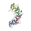
| ||||||||
| Unit cell |
|
-Components
| #1: Protein | Mass: 7999.139 Da / Num. of mol.: 4 Source method: isolated from a genetically manipulated source Source: (gene. exp.) Streptococcus phage C1 (virus) / Gene: orf9 / Production host:  Escherichia coli (E. coli) / References: UniProt: Q7Y3F3 Escherichia coli (E. coli) / References: UniProt: Q7Y3F3#2: Chemical | ChemComp-MRD / ( | 2-Methyl-2,4-pentanediol  Mass: 118.174 Da / Num. of mol.: 1 / Source method: obtained synthetically / Formula: C6H14O2 / Comment: precipitant*YM Mass: 118.174 Da / Num. of mol.: 1 / Source method: obtained synthetically / Formula: C6H14O2 / Comment: precipitant*YM#3: Chemical | ChemComp-MPD / ( | 2-Methyl-2,4-pentanediol  Mass: 118.174 Da / Num. of mol.: 1 / Source method: obtained synthetically / Formula: C6H14O2 / Comment: precipitant*YM Mass: 118.174 Da / Num. of mol.: 1 / Source method: obtained synthetically / Formula: C6H14O2 / Comment: precipitant*YM#4: Water | ChemComp-HOH / | Water Mass: 18.015 Da / Num. of mol.: 373 / Source method: isolated from a natural source / Formula: H2O Mass: 18.015 Da / Num. of mol.: 373 / Source method: isolated from a natural source / Formula: H2O |
|---|
-Experimental details
-Experiment
| Experiment | Method: X-RAY DIFFRACTION / Number of used crystals: 1 |
|---|
- Sample preparation
| Crystal | Density Matthews: 2.93 Å3/Da / Density % sol: 57.95 % |
|---|---|
| Crystal grow | Method: vapor diffusion, hanging drop / pH: 6 Details: 25% MPD, 0.1 M Hepes, 0.2 M Sodium citrate, pH 6.0, VAPOR DIFFUSION, HANGING DROP |
-Data collection
| Diffraction |
| ||||||||||||||||||
|---|---|---|---|---|---|---|---|---|---|---|---|---|---|---|---|---|---|---|---|
| Diffraction source |
| ||||||||||||||||||
| Detector |
| ||||||||||||||||||
| Radiation |
| ||||||||||||||||||
| Radiation wavelength |
| ||||||||||||||||||
| Reflection | Resolution: 1.4→30 Å / Num. obs: 71861 / % possible obs: 98.4 % / Redundancy: 7.1 % / Biso Wilson estimate: 20.45 Å2 / Rmerge(I) obs: 0.056 | ||||||||||||||||||
| Reflection shell | Resolution: 1.4→1.45 Å / Redundancy: 6.8 % / Rmerge(I) obs: 0.58 / % possible all: 100 |

- Processing
| Software |
| |||||||||||||||||||||||||||||||||||||||||||||||||||||||||||||||||||||||||||||||||||||||||||||||||||||||||||||||||||||||||||||
|---|---|---|---|---|---|---|---|---|---|---|---|---|---|---|---|---|---|---|---|---|---|---|---|---|---|---|---|---|---|---|---|---|---|---|---|---|---|---|---|---|---|---|---|---|---|---|---|---|---|---|---|---|---|---|---|---|---|---|---|---|---|---|---|---|---|---|---|---|---|---|---|---|---|---|---|---|---|---|---|---|---|---|---|---|---|---|---|---|---|---|---|---|---|---|---|---|---|---|---|---|---|---|---|---|---|---|---|---|---|---|---|---|---|---|---|---|---|---|---|---|---|---|---|---|---|---|
| Refinement | Method to determine structure: S-SAD / Resolution: 1.4→28 Å / Cor.coef. Fo:Fc: 0.9516 / Cor.coef. Fo:Fc free: 0.9533 / Cross valid method: THROUGHOUT / σ(F): 0
| |||||||||||||||||||||||||||||||||||||||||||||||||||||||||||||||||||||||||||||||||||||||||||||||||||||||||||||||||||||||||||||
| Displacement parameters | Biso mean: 25.55 Å2
| |||||||||||||||||||||||||||||||||||||||||||||||||||||||||||||||||||||||||||||||||||||||||||||||||||||||||||||||||||||||||||||
| Refine analyze | Luzzati coordinate error obs: 0.179 Å | |||||||||||||||||||||||||||||||||||||||||||||||||||||||||||||||||||||||||||||||||||||||||||||||||||||||||||||||||||||||||||||
| Refinement step | Cycle: LAST / Resolution: 1.4→28 Å
| |||||||||||||||||||||||||||||||||||||||||||||||||||||||||||||||||||||||||||||||||||||||||||||||||||||||||||||||||||||||||||||
| Refine LS restraints |
| |||||||||||||||||||||||||||||||||||||||||||||||||||||||||||||||||||||||||||||||||||||||||||||||||||||||||||||||||||||||||||||
| LS refinement shell | Resolution: 1.4→1.44 Å / Total num. of bins used: 20
| |||||||||||||||||||||||||||||||||||||||||||||||||||||||||||||||||||||||||||||||||||||||||||||||||||||||||||||||||||||||||||||
| Refinement TLS params. | Method: refined / Refine-ID: X-RAY DIFFRACTION
| |||||||||||||||||||||||||||||||||||||||||||||||||||||||||||||||||||||||||||||||||||||||||||||||||||||||||||||||||||||||||||||
| Refinement TLS group |
|
