 Movie
Movie Controller


+ Open data

- Basic information
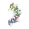
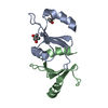
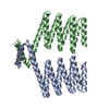
| Entry | Database: PDB / ID: 4zrz | ||||||
|---|---|---|---|---|---|---|---|
| Title | PlyCB mutant R66E | ||||||
 Components Components | PlyCB | ||||||
 Keywords Keywords |  Antimicrobial protein / Viral protein / bacteriocidal / bacteriophage lysin / cell-binding subunit / octameric / cell-wall-cutting Antimicrobial protein / Viral protein / bacteriocidal / bacteriophage lysin / cell-binding subunit / octameric / cell-wall-cutting | ||||||
| Function / homology | Signal recognition particle alu RNA binding heterodimer, srp9/1 - #190 / : / Streptococcus virus C1, PlyCB / Signal recognition particle alu RNA binding heterodimer, srp9/1 / killing of cells of another organism / 2-Layer Sandwich / identical protein binding / Alpha Beta / Endolysin PlyC, small cell-wall binding subunit Function and homology information Function and homology information | ||||||
| Biological species |  Streptococcus phage C1 (virus) Streptococcus phage C1 (virus) | ||||||
| Method | X-RAY DIFFRACTION / MOLECULAR REPLACEMENT / Resolution: 1.72 Å | ||||||
 Authors Authors | Gallagher, D.T. / Nelson, D.C. / Shen, Y. | ||||||
 Citation Citation | Journal: Elife / Year: 2016 Title: A bacteriophage endolysin that eliminates intracellular streptococci. Authors: Shen, Y. / Barros, M. / Vennemann, T. / Gallagher, D.T. / Yin, Y. / Linden, S.B. / Heselpoth, R.D. / Spencer, D.J. / Donovan, D.M. / Moult, J. / Fischetti, V.A. / Heinrich, F. / Losche, M. / Nelson, D.C. | ||||||
| History |
|
- Structure visualization
| Structure viewer | Molecule: MolmilJmol/JSmol |
|---|
- Downloads & links
-Download
| PDBx/mmCIF format | 4zrz.cif.gz | 40.2 KB | Display | PDBx/mmCIF format |
|---|---|---|---|---|
| PDB format | pdb4zrz.ent.gz | 27.8 KB | Display | PDB format |
| PDBx/mmJSON format | 4zrz.json.gz | Tree view | PDBx/mmJSON format | |
| Others |  Other downloads Other downloads |
-Validation report
| Arichive directory | https://data.pdbj.org/pub/pdb/validation_reports/zr/4zrzftp://data.pdbj.org/pub/pdb/validation_reports/zr/4zrz | HTTPS FTP |
|---|
-Related structure data
| Related structure data |  4f87S S: Starting model for refinement |
|---|---|
| Similar structure data |


-Links
 PDBj
PDBj- Assembly
| Deposited unit | 
| ||||||||
|---|---|---|---|---|---|---|---|---|---|
| 1 | 
| ||||||||
| Unit cell |
| ||||||||
| Components on special symmetry positions |
|
-Components
| #1: Protein | Mass: 7839.863 Da / Num. of mol.: 2 Source method: isolated from a genetically manipulated source Source: (gene. exp.) Streptococcus phage C1 (virus) / Gene: orf9 / Plasmid: pET15b / Production host:  Escherichia coli (E. coli) / Strain (production host): BL21(DE3) / References: UniProt: Q7Y3F3 Escherichia coli (E. coli) / Strain (production host): BL21(DE3) / References: UniProt: Q7Y3F3#2: Water | ChemComp-HOH / | Water Mass: 18.015 Da / Num. of mol.: 105 / Source method: isolated from a natural source / Formula: H2O Mass: 18.015 Da / Num. of mol.: 105 / Source method: isolated from a natural source / Formula: H2O |
|---|
-Experimental details
-Experiment
| Experiment | Method: X-RAY DIFFRACTION / Number of used crystals: 1 |
|---|
- Sample preparation
| Crystal | Density Matthews: 2.95 Å3/Da / Density % sol: 58.28 % |
|---|---|
| Crystal grow | Temperature: 298 K / Method: vapor diffusion, sitting drop / pH: 6 / Details: 14 mg/mL ptn, 43% MPD, 30 mM AmSO4, 70 mM Na Hepes |
-Data collection
| Diffraction | Mean temperature: 100 K | |||||||||||||||||||||||||||||||||||||||||||||||||||||||||||||||||||||||||||||||||||||||||||||||||||
|---|---|---|---|---|---|---|---|---|---|---|---|---|---|---|---|---|---|---|---|---|---|---|---|---|---|---|---|---|---|---|---|---|---|---|---|---|---|---|---|---|---|---|---|---|---|---|---|---|---|---|---|---|---|---|---|---|---|---|---|---|---|---|---|---|---|---|---|---|---|---|---|---|---|---|---|---|---|---|---|---|---|---|---|---|---|---|---|---|---|---|---|---|---|---|---|---|---|---|---|---|
| Diffraction source | Source: ROTATING ANODE / Type: RIGAKU MICROMAX-007 HF / Wavelength: 1.54 Å | |||||||||||||||||||||||||||||||||||||||||||||||||||||||||||||||||||||||||||||||||||||||||||||||||||
| Detector | Type: RIGAKU RAXIS IV++ / Detector: IMAGE PLATE / Date: Nov 2, 2012 | |||||||||||||||||||||||||||||||||||||||||||||||||||||||||||||||||||||||||||||||||||||||||||||||||||
| Radiation | Protocol: SINGLE WAVELENGTH / Monochromatic (M) / Laue (L): M / Scattering type: x-ray | |||||||||||||||||||||||||||||||||||||||||||||||||||||||||||||||||||||||||||||||||||||||||||||||||||
| Radiation wavelength | Wavelength: 1.54 Å / Relative weight: 1 | |||||||||||||||||||||||||||||||||||||||||||||||||||||||||||||||||||||||||||||||||||||||||||||||||||
| Reflection | Resolution: 1.72→19.66 Å / Num. obs: 20025 / % possible obs: 98.1 % / Redundancy: 5.18 % / Rmerge(I) obs: 0.028 / Χ2: 0.98 / Net I/σ(I): 25.4 / Num. measured all: 104605 / Scaling rejects: 785 | |||||||||||||||||||||||||||||||||||||||||||||||||||||||||||||||||||||||||||||||||||||||||||||||||||
| Reflection shell | Diffraction-ID: 1
|
- Processing
| Software |
| |||||||||||||||||||||||||||||||||||||||||||||||||||||||||||||||||||||||||||
|---|---|---|---|---|---|---|---|---|---|---|---|---|---|---|---|---|---|---|---|---|---|---|---|---|---|---|---|---|---|---|---|---|---|---|---|---|---|---|---|---|---|---|---|---|---|---|---|---|---|---|---|---|---|---|---|---|---|---|---|---|---|---|---|---|---|---|---|---|---|---|---|---|---|---|---|---|
| Refinement | Method to determine structure: MOLECULAR REPLACEMENT Starting model: 4F87 Resolution: 1.72→14 Å / Cor.coef. Fo:Fc: 0.956 / Cor.coef. Fo:Fc free: 0.918 / WRfactor Rfree: 0.2492 / WRfactor Rwork: 0.2174 / FOM work R set: 0.8182 / SU B: 2.181 / SU ML: 0.071 / SU R Cruickshank DPI: 0.1022 / SU Rfree: 0.1034 / Cross valid method: THROUGHOUT / σ(F): 0 / ESU R: 0.102 / ESU R Free: 0.103 / Stereochemistry target values: MAXIMUM LIKELIHOOD Details: HYDROGENS HAVE BEEN ADDED IN THE RIDING POSITIONS U VALUES : REFINED INDIVIDUALLY
| |||||||||||||||||||||||||||||||||||||||||||||||||||||||||||||||||||||||||||
| Solvent computation | Ion probe radii: 0.8 Å / Shrinkage radii: 0.8 Å / VDW probe radii: 1.2 Å / Solvent model: MASK | |||||||||||||||||||||||||||||||||||||||||||||||||||||||||||||||||||||||||||
| Displacement parameters | Biso max: 82.67 Å2 / Biso mean: 33.254 Å2 / Biso min: 19 Å2
| |||||||||||||||||||||||||||||||||||||||||||||||||||||||||||||||||||||||||||
| Refinement step | Cycle: final / Resolution: 1.72→14 Å
| |||||||||||||||||||||||||||||||||||||||||||||||||||||||||||||||||||||||||||
| Refine LS restraints |
| |||||||||||||||||||||||||||||||||||||||||||||||||||||||||||||||||||||||||||
| LS refinement shell | Resolution: 1.72→1.764 Å / Total num. of bins used: 20
|
