 Movie
Movie Controller

[English] 日本語
 Yorodumi
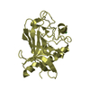
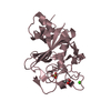
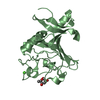
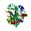
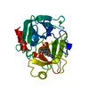
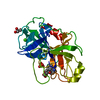
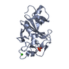
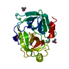
- PDB-4esq: Crystal structure of the extracellular domain of PknH from Mycoba... -
+ Open data

- Basic information
| Entry | Database: PDB / ID: 4esq | ||||||
|---|---|---|---|---|---|---|---|
| Title | Crystal structure of the extracellular domain of PknH from Mycobacterium tuberculosis | ||||||
 Components Components | Serine/threonine protein kinase Serine/threonine-specific protein kinase Serine/threonine-specific protein kinase | ||||||
 Keywords Keywords | TRANSFERASE / Receptor Kinase / Membrane | ||||||
| Function / homology |  Function and homology information Function and homology informationregulation of lipid biosynthetic process / negative regulation of growth / negative regulation of catalytic activity / response to host immune response / cell wall / positive regulation of catalytic activity / plasma membrane => GO:0005886 / positive regulation of DNA binding / protein autophosphorylation / membrane => GO:0016020 ...regulation of lipid biosynthetic process / negative regulation of growth / negative regulation of catalytic activity / response to host immune response / cell wall / positive regulation of catalytic activity / plasma membrane => GO:0005886 / positive regulation of DNA binding / protein autophosphorylation / membrane => GO:0016020 / non-specific serine/threonine protein kinase / protein kinase activity / protein serine/threonine kinase activity / positive regulation of DNA-templated transcription / ATP binding / plasma membraneSimilarity search - Function | ||||||
| Biological species |   Mycobacterium tuberculosis (bacteria) Mycobacterium tuberculosis (bacteria) | ||||||
| Method | X-RAY DIFFRACTION / SYNCHROTRON / SAD / Resolution: 1.7 Å | ||||||
 Authors Authors | Cavazos, A. / Prigozhin, D.M. / Alber, T. | ||||||
 Citation Citation | Journal: J.Mol.Biol. / Year: 2012 Title: Structure of the sensor domain of Mycobacterium tuberculosis PknH receptor kinase reveals a conserved binding cleft. Authors: Cavazos, A. / Prigozhin, D.M. / Alber, T. | ||||||
| History |
|
- Structure visualization
| Structure viewer | Molecule: MolmilJmol/JSmol |
|---|
- Downloads & links
-Download
| PDBx/mmCIF format | 4esq.cif.gz | 53.6 KB | Display | PDBx/mmCIF format |
|---|---|---|---|---|
| PDB format | pdb4esq.ent.gz | 39.6 KB | Display | PDB format |
| PDBx/mmJSON format | 4esq.json.gz | Tree view | PDBx/mmJSON format | |
| Others |  Other downloads Other downloads |
-Validation report
| Arichive directory | https://data.pdbj.org/pub/pdb/validation_reports/es/4esqftp://data.pdbj.org/pub/pdb/validation_reports/es/4esq | HTTPS FTP |
|---|
-Related structure data
| Similar structure data |
|---|


-Links
 PDBj
PDBj- Assembly
| Deposited unit | 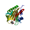
| ||||||||
|---|---|---|---|---|---|---|---|---|---|
| 1 |
| ||||||||
| Unit cell |
|
-Components
| #1: Protein | Serine/threonine-specific protein kinase Mass: 20661.994 Da / Num. of mol.: 1 / Fragment: unp residues 83332-83523 Source method: isolated from a genetically manipulated source Source: (gene. exp.) Mycobacterium tuberculosis (bacteria) / Strain: H37Rv / Gene: pknH, Rv1266c / Production host: Escherichia coli (E. coli) / Strain (production host): BL21 (DE3) CodonPlus / References: UniProt: A5U1W4, UniProt: P9WI71*PLUS | ||
|---|---|---|---|
| #2: Chemical | ChemComp-BTB / Bis-tris methane  Mass: 209.240 Da / Num. of mol.: 1 / Source method: obtained synthetically / Formula: C8H19NO5 / Comment: pH buffer*YM Mass: 209.240 Da / Num. of mol.: 1 / Source method: obtained synthetically / Formula: C8H19NO5 / Comment: pH buffer*YM | ||
| #3: Chemical |   Mass: 158.925 Da / Num. of mol.: 2 / Source method: obtained synthetically / Formula: Tb Mass: 158.925 Da / Num. of mol.: 2 / Source method: obtained synthetically / Formula: Tb#4: Water | ChemComp-HOH / | Water Mass: 18.015 Da / Num. of mol.: 221 / Source method: isolated from a natural source / Formula: H2O Mass: 18.015 Da / Num. of mol.: 221 / Source method: isolated from a natural source / Formula: H2O |
-Experimental details
-Experiment
| Experiment | Method: X-RAY DIFFRACTION / Number of used crystals: 1 |
|---|
- Sample preparation
| Crystal | Density Matthews: 2.01 Å3/Da / Density % sol: 38.88 % |
|---|---|
| Crystal grow | Temperature: 291 K / Method: vapor diffusion / pH: 5.5 Details: 0.1 M Bis-Tris, 0.2 M ammonium acetate, 25% PEG 3350, 50mM terbium nitrate, pH 5.5, VAPOR DIFFUSION, temperature 291K |
-Data collection
| Diffraction | Mean temperature: 100 K | |||||||||||||||||||||||||||||||||||||||||||||||||||||||||||||||||||||||||||||
|---|---|---|---|---|---|---|---|---|---|---|---|---|---|---|---|---|---|---|---|---|---|---|---|---|---|---|---|---|---|---|---|---|---|---|---|---|---|---|---|---|---|---|---|---|---|---|---|---|---|---|---|---|---|---|---|---|---|---|---|---|---|---|---|---|---|---|---|---|---|---|---|---|---|---|---|---|---|---|
| Diffraction source | Source: SYNCHROTRON / Site: ALS  / Beamline: 8.3.1 / Wavelength: 1.1159 Å / Beamline: 8.3.1 / Wavelength: 1.1159 Å | |||||||||||||||||||||||||||||||||||||||||||||||||||||||||||||||||||||||||||||
| Detector | Type: ADSC QUANTUM 315r / Detector: CCD / Date: Sep 27, 2011 | |||||||||||||||||||||||||||||||||||||||||||||||||||||||||||||||||||||||||||||
| Radiation | Monochromator: Double flat crystal, Si(111) / Protocol: SINGLE WAVELENGTH / Monochromatic (M) / Laue (L): M / Scattering type: x-ray | |||||||||||||||||||||||||||||||||||||||||||||||||||||||||||||||||||||||||||||
| Radiation wavelength | Wavelength: 1.1159 Å / Relative weight: 1 | |||||||||||||||||||||||||||||||||||||||||||||||||||||||||||||||||||||||||||||
| Reflection | Redundancy: 3.8 % / Av σ(I) over netI: 17.44 / Number: 69365 / Rmerge(I) obs: 0.061 / Χ2: 1.01 / D res high: 1.7 Å / D res low: 50 Å / Num. obs: 18089 / % possible obs: 98.8 | |||||||||||||||||||||||||||||||||||||||||||||||||||||||||||||||||||||||||||||
| Diffraction reflection shell |
| |||||||||||||||||||||||||||||||||||||||||||||||||||||||||||||||||||||||||||||
| Reflection | Resolution: 1.7→50 Å / Num. obs: 34200 / % possible obs: 98.8 % / Redundancy: 3.8 % / Rmerge(I) obs: 0.061 / Χ2: 1.015 / Net I/σ(I): 17.1 | |||||||||||||||||||||||||||||||||||||||||||||||||||||||||||||||||||||||||||||
| Reflection shell |
|
-Phasing
| Phasing | Method: SAD | ||||||||||||||||||||||||||||||||||||||||||
|---|---|---|---|---|---|---|---|---|---|---|---|---|---|---|---|---|---|---|---|---|---|---|---|---|---|---|---|---|---|---|---|---|---|---|---|---|---|---|---|---|---|---|---|
| Phasing dm | FOM : 0.69 / FOM acentric: 0.71 / FOM centric: 0.51 / Reflection: 8491 / Reflection acentric: 7763 / Reflection centric: 728 | ||||||||||||||||||||||||||||||||||||||||||
| Phasing dm shell |
|
- Processing
| Software |
| |||||||||||||||||||||||||||||||||||||||||||||||||||||||||||||||||||||||||||||||||||||||||||
|---|---|---|---|---|---|---|---|---|---|---|---|---|---|---|---|---|---|---|---|---|---|---|---|---|---|---|---|---|---|---|---|---|---|---|---|---|---|---|---|---|---|---|---|---|---|---|---|---|---|---|---|---|---|---|---|---|---|---|---|---|---|---|---|---|---|---|---|---|---|---|---|---|---|---|---|---|---|---|---|---|---|---|---|---|---|---|---|---|---|---|---|---|
| Refinement | Method to determine structure: SAD / Resolution: 1.7→48.784 Å / Occupancy max: 1 / Occupancy min: 0.23 / SU ML: 0.39 / σ(F): 1.35 / Phase error: 19.09 / Stereochemistry target values: ML
| |||||||||||||||||||||||||||||||||||||||||||||||||||||||||||||||||||||||||||||||||||||||||||
| Solvent computation | Shrinkage radii: 0.83 Å / VDW probe radii: 1.1 Å / Solvent model: FLAT BULK SOLVENT MODEL / Bsol: 28.934 Å2 / ksol: 0.317 e/Å3 | |||||||||||||||||||||||||||||||||||||||||||||||||||||||||||||||||||||||||||||||||||||||||||
| Displacement parameters | Biso max: 55.6 Å2 / Biso mean: 18.0416 Å2 / Biso min: 7.58 Å2
| |||||||||||||||||||||||||||||||||||||||||||||||||||||||||||||||||||||||||||||||||||||||||||
| Refinement step | Cycle: LAST / Resolution: 1.7→48.784 Å
| |||||||||||||||||||||||||||||||||||||||||||||||||||||||||||||||||||||||||||||||||||||||||||
| Refine LS restraints |
| |||||||||||||||||||||||||||||||||||||||||||||||||||||||||||||||||||||||||||||||||||||||||||
| LS refinement shell | Refine-ID: X-RAY DIFFRACTION / Total num. of bins used: 12
|
