 Movie
Movie Controller

[English] 日本語
 Yorodumi
- PDB-4erm: Crystal structure of the dATP inhibited E. coli class Ia ribonucl... -
+ Open data

- Basic information
| Entry | Database: PDB / ID: 4erm | ||||||
|---|---|---|---|---|---|---|---|
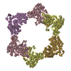
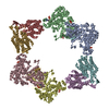
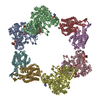
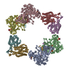
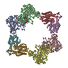
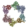
| Title | Crystal structure of the dATP inhibited E. coli class Ia ribonucleotide reductase complex at 4 Angstroms resolution | ||||||
 Components Components | (Ribonucleoside-diphosphate reductase 1 subunit ...) x 2 | ||||||
 Keywords Keywords | OXIDOREDUCTASE / protein-protein complex / alpha/beta barrel / atp cone / di-iron center / RNR alpha / RNR beta / thioredoxin / Ribonucleotide reduction / Cytosol | ||||||
| Function / homology |  Function and homology information Function and homology informationribonucleoside diphosphate metabolic process / 2'-deoxyribonucleotide biosynthetic process / ribonucleoside-diphosphate reductase complex / nucleobase-containing small molecule interconversion / ribonucleoside-diphosphate reductase / ribonucleoside-diphosphate reductase activity, thioredoxin disulfide as acceptor / deoxyribonucleotide biosynthetic process / protein folding chaperone / iron ion binding / ATP binding ...ribonucleoside diphosphate metabolic process / 2'-deoxyribonucleotide biosynthetic process / ribonucleoside-diphosphate reductase complex / nucleobase-containing small molecule interconversion / ribonucleoside-diphosphate reductase / ribonucleoside-diphosphate reductase activity, thioredoxin disulfide as acceptor / deoxyribonucleotide biosynthetic process / protein folding chaperone / iron ion binding / ATP binding / identical protein binding / cytoplasm / cytosol Similarity search - Function | ||||||
| Biological species |  | ||||||
| Method |  X-RAY DIFFRACTION / SYNCHROTRON / FOURIER SYNTHESIS / Resolution: 3.95 Å X-RAY DIFFRACTION / SYNCHROTRON / FOURIER SYNTHESIS / Resolution: 3.95 Å | ||||||
 Authors Authors | Zimanyi, C.M. / Drennan, C.L. | ||||||
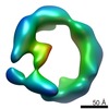
 Citation Citation | Journal: Structure / Year: 2012 Title: Tangled up in knots: structures of inactivated forms of E. coli class Ia ribonucleotide reductase. Authors: Christina M Zimanyi / Nozomi Ando / Edward J Brignole / Francisco J Asturias / Joanne Stubbe / Catherine L Drennan /  Abstract: Ribonucleotide reductases (RNRs) provide the precursors for DNA biosynthesis and repair and are successful targets for anticancer drugs such as clofarabine and gemcitabine. Recently, we reported that ...Ribonucleotide reductases (RNRs) provide the precursors for DNA biosynthesis and repair and are successful targets for anticancer drugs such as clofarabine and gemcitabine. Recently, we reported that dATP inhibits E. coli class Ia RNR by driving formation of RNR subunits into α4β4 rings. Here, we present the first X-ray structure of a gemcitabine-inhibited E. coli RNR and show that the previously described α4β4 rings can interlock to form an unprecedented (α4β4)2 megacomplex. This complex is also seen in a higher-resolution dATP-inhibited RNR structure presented here, which employs a distinct crystal lattice from that observed in the gemcitabine-inhibited case. With few reported examples of protein catenanes, we use data from small-angle X-ray scattering and electron microscopy to both understand the solution conditions that contribute to concatenation in RNRs as well as present a mechanism for the formation of these unusual structures. | ||||||
| History |
|
- Structure visualization
| Structure viewer | Molecule: MolmilJmol/JSmol |
|---|
- Downloads & links
-Download
| PDBx/mmCIF format | 4erm.cif.gz | 776.4 KB | Display | PDBx/mmCIF format |
|---|---|---|---|---|
| PDB format | pdb4erm.ent.gz | 655.1 KB | Display | PDB format |
| PDBx/mmJSON format | 4erm.json.gz | Tree view | PDBx/mmJSON format | |
| Others |  Other downloads Other downloads |
-Validation report
| Summary document | 4erm_validation.pdf.gz | 3.7 MB | Display | wwPDB validaton report |
|---|---|---|---|---|
| Full document | 4erm_full_validation.pdf.gz | 3.9 MB | Display | |
| Data in XML | 4erm_validation.xml.gz | 180.5 KB | Display | |
| Data in CIF | 4erm_validation.cif.gz | 234.7 KB | Display | |
| Arichive directory | https://data.pdbj.org/pub/pdb/validation_reports/er/4ermftp://data.pdbj.org/pub/pdb/validation_reports/er/4erm | HTTPS FTP |
-Related structure data
| Related structure data |  5430C  5431C  5432C  5433C  5434C  5435C  5436C  5437C 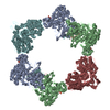 4erpC  3uusS S: Starting model for refinement C: citing same article ( |
|---|---|
| Similar structure data |

-Links
 PDBj
PDBj


- Assembly
| Deposited unit | 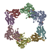
| ||||||||
|---|---|---|---|---|---|---|---|---|---|
| 1 |
| ||||||||
| Unit cell |
|
-Components
-Ribonucleoside-diphosphate reductase 1 subunit ... , 2 types, 8 molecules ABCDEFGH
| #1: Protein | Mass: 85877.086 Da / Num. of mol.: 4 Source method: isolated from a genetically manipulated source Source: (gene. exp.) References: UniProt: P00452, ribonucleoside-diphosphate reductase #2: Protein | Mass: 43426.863 Da / Num. of mol.: 4 Source method: isolated from a genetically manipulated source Source: (gene. exp.) References: UniProt: P69924, ribonucleoside-diphosphate reductase |
|---|
-Non-polymers , 5 types, 28 molecules 


| #3: Chemical | ChemComp-DTP /  Mass: 491.182 Da / Num. of mol.: 8 / Source method: obtained synthetically / Formula: C10H16N5O12P3 Mass: 491.182 Da / Num. of mol.: 8 / Source method: obtained synthetically / Formula: C10H16N5O12P3#4: Chemical | ChemComp-DAT /  Mass: 411.202 Da / Num. of mol.: 4 / Source method: obtained synthetically / Formula: C10H15N5O9P2 Mass: 411.202 Da / Num. of mol.: 4 / Source method: obtained synthetically / Formula: C10H15N5O9P2#5: Chemical | ChemComp-MG /  Mass: 24.305 Da / Num. of mol.: 4 / Source method: obtained synthetically / Formula: Mg Mass: 24.305 Da / Num. of mol.: 4 / Source method: obtained synthetically / Formula: Mg#6: Chemical | ChemComp-FEO /  Mass: 127.689 Da / Num. of mol.: 4 / Source method: obtained synthetically / Formula: Fe2O Mass: 127.689 Da / Num. of mol.: 4 / Source method: obtained synthetically / Formula: Fe2O#7: Water | ChemComp-HOH / | Mass: 18.015 Da / Num. of mol.: 8 / Source method: isolated from a natural source / Formula: H2O |
|---|
-Experimental details
-Experiment
| Experiment | Method: X-RAY DIFFRACTION / Number of used crystals: 1 |
|---|
- Sample preparation
| Crystal | Density Matthews: 3.08 Å3/Da / Density % sol: 60.07 % |
|---|---|
| Crystal grow | Temperature: 298 K / Method: vapor diffusion, hanging drop / pH: 7.5 Details: Precipitant conditions: 10% PEG 3350, 0.1 M MOPS pH 7.5, 0.2 M Magnesium Acetate, 0.025 M Magnesium Chloride, 0.006 M n-Nonyl-beta-D-maltopyranoside, and 5% glycerol. Mixed 1:1 with protein. ...Details: Precipitant conditions: 10% PEG 3350, 0.1 M MOPS pH 7.5, 0.2 M Magnesium Acetate, 0.025 M Magnesium Chloride, 0.006 M n-Nonyl-beta-D-maltopyranoside, and 5% glycerol. Mixed 1:1 with protein., VAPOR DIFFUSION, HANGING DROP, temperature 298K |
-Data collection
| Diffraction | Mean temperature: 100 K |
|---|---|
| Diffraction source | Source: SYNCHROTRON / Site: APS / Beamline: 24-ID-C / Wavelength: 0.9794 Å |
| Detector | Type: ADSC QUANTUM 315 / Detector: CCD / Date: Feb 25, 2011 |
| Radiation | Monochromator: Si(111) / Protocol: SINGLE WAVELENGTH / Monochromatic (M) / Laue (L): M / Scattering type: x-ray |
| Radiation wavelength | Wavelength: 0.9794 Å / Relative weight: 1 |
| Reflection | Resolution: 3.95→30 Å / Num. all: 55058 / Num. obs: 53173 / % possible obs: 96.6 % / Observed criterion σ(F): 0 / Observed criterion σ(I): 0 / Redundancy: 2.9 % / Rsym value: 0.161 / Net I/σ(I): 5 |
| Reflection shell | Resolution: 3.95→4.09 Å / Redundancy: 2.9 % / Mean I/σ(I) obs: 2.3 / Num. unique all: 5306 / Rsym value: 0.457 / % possible all: 96.5 |
- Processing
| Software |
| |||||||||||||||||||||||||
|---|---|---|---|---|---|---|---|---|---|---|---|---|---|---|---|---|---|---|---|---|---|---|---|---|---|---|
| Refinement | Method to determine structure: FOURIER SYNTHESIS Starting model: 3UUS Resolution: 3.95→30 Å / σ(F): 0 / Stereochemistry target values: Engh & Huber
| |||||||||||||||||||||||||
| Refinement step | Cycle: LAST / Resolution: 3.95→30 Å
| |||||||||||||||||||||||||
| Refine LS restraints |
| |||||||||||||||||||||||||
| LS refinement shell | Resolution: 3.95→4.09 Å
|
