 Movie
Movie Controller

[English] 日本語
 Yorodumi
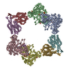
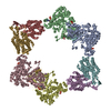
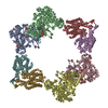
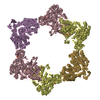
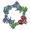
- PDB-5cnv: Crystal structure of the dATP inhibited E. coli class Ia ribonucl... -
+ Open data

- Basic information
| Entry | Database: PDB / ID: 5cnv | ||||||||||||
|---|---|---|---|---|---|---|---|---|---|---|---|---|---|
| Title | Crystal structure of the dATP inhibited E. coli class Ia ribonucleotide reductase complex bound to GDP and TTP at 3.20 Angstroms resolution | ||||||||||||
 Components Components | (Ribonucleoside-diphosphate reductase 1 subunit ... Ribonucleotide reductase) x 2 Ribonucleotide reductase) x 2 | ||||||||||||
 Keywords Keywords | OXIDOREDUCTASE / allostery / substrate specificity / ribonucleotide reductase / nucleotide metabolism | ||||||||||||
| Function / homology |  Function and homology information Function and homology informationribonucleoside diphosphate metabolic process / 2'-deoxyribonucleotide biosynthetic process / nucleobase-containing small molecule interconversion / ribonucleoside-diphosphate reductase complex / ribonucleoside-diphosphate reductase / ribonucleoside-diphosphate reductase activity, thioredoxin disulfide as acceptor / deoxyribonucleotide biosynthetic process / protein folding chaperone / DNA replication / iron ion binding ...ribonucleoside diphosphate metabolic process / 2'-deoxyribonucleotide biosynthetic process / nucleobase-containing small molecule interconversion / ribonucleoside-diphosphate reductase complex / ribonucleoside-diphosphate reductase / ribonucleoside-diphosphate reductase activity, thioredoxin disulfide as acceptor / deoxyribonucleotide biosynthetic process / protein folding chaperone / DNA replication / iron ion binding / ATP binding / identical protein binding / cytosol / cytoplasmSimilarity search - Function | ||||||||||||
| Biological species |  Escherichia coli (E. coli) Escherichia coli (E. coli) | ||||||||||||
| Method | X-RAY DIFFRACTION / SYNCHROTRON / MOLECULAR REPLACEMENT / Resolution: 3.2 Å | ||||||||||||
 Authors Authors | Chen, P.Y.-T. / Zimanyi, C.M. / Funk, M.A. / Drennan, C.L. | ||||||||||||
| Funding support |  United States, 3items United States, 3items
| ||||||||||||
 Citation Citation | Journal: Elife / Year: 2016 Title: Molecular basis for allosteric specificity regulation in class Ia ribonucleotide reductase from Escherichia coli. Authors: Zimanyi, C.M. / Chen, P.Y. / Kang, G. / Funk, M.A. / Drennan, C.L. | ||||||||||||
| History |
|
- Structure visualization
| Structure viewer | Molecule: MolmilJmol/JSmol |
|---|
- Downloads & links
-Download
| PDBx/mmCIF format | 5cnv.cif.gz | 858 KB | Display | PDBx/mmCIF format |
|---|---|---|---|---|
| PDB format | pdb5cnv.ent.gz | 707.2 KB | Display | PDB format |
| PDBx/mmJSON format | 5cnv.json.gz | Tree view | PDBx/mmJSON format | |
| Others |  Other downloads Other downloads |
-Validation report
| Arichive directory | https://data.pdbj.org/pub/pdb/validation_reports/cn/5cnvftp://data.pdbj.org/pub/pdb/validation_reports/cn/5cnv | HTTPS FTP |
|---|
-Related structure data
| Related structure data |  5cnsC 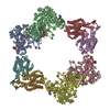 5cntC  5cnuC 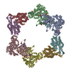 4ermS C: citing same article ( S: Starting model for refinement |
|---|---|
| Similar structure data |


-Links
 PDBj
PDBj
- Assembly
| Deposited unit | 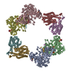
| ||||||||
|---|---|---|---|---|---|---|---|---|---|
| 1 |
| ||||||||
| Unit cell |
|
-Components
-Ribonucleoside-diphosphate reductase 1 subunit ... , 2 types, 8 molecules ABCDEFGH
| #1: Protein | Ribonucleotide reductase / Protein B1 / Ribonucleoside-diphosphate reductase 1 R1 subunit / Ribonucleotide reductase 1 Mass: 85877.086 Da / Num. of mol.: 4 Source method: isolated from a genetically manipulated source Source: (gene. exp.) Escherichia coli (strain K12) (bacteria)Strain: K12 / Gene: nrdA, dnaF, b2234, JW2228 / Production host: Escherichia coli (E. coli)References: UniProt: P00452, ribonucleoside-diphosphate reductase#2: Protein | Ribonucleotide reductase / Protein B2 / Protein R2 / Ribonucleotide reductase 1Mass: 43426.863 Da / Num. of mol.: 4 Source method: isolated from a genetically manipulated source Source: (gene. exp.) Escherichia coli (strain K12) (bacteria)Strain: K12 / Gene: nrdB, ftsB, b2235, JW2229 / Production host: Escherichia coli (E. coli)References: UniProt: P69924, ribonucleoside-diphosphate reductase |
|---|
-Non-polymers , 6 types, 89 molecules 


| #3: Chemical | ChemComp-GDP / Guanosine diphosphate Type: RNA linking / Mass: 443.201 Da / Num. of mol.: 4 / Source method: obtained synthetically / Formula: C10H15N5O11P2 / Comment: GDP, energy-carrying molecule*YM Type: RNA linking / Mass: 443.201 Da / Num. of mol.: 4 / Source method: obtained synthetically / Formula: C10H15N5O11P2 / Comment: GDP, energy-carrying molecule*YM#4: Chemical | ChemComp-DAT / Deoxyadenosine diphosphate Mass: 411.202 Da / Num. of mol.: 4 / Source method: obtained synthetically / Formula: C10H15N5O9P2 Mass: 411.202 Da / Num. of mol.: 4 / Source method: obtained synthetically / Formula: C10H15N5O9P2#5: Chemical | ChemComp-MG /  Mass: 24.305 Da / Num. of mol.: 8 / Source method: obtained synthetically / Formula: Mg Mass: 24.305 Da / Num. of mol.: 8 / Source method: obtained synthetically / Formula: Mg#6: Chemical | ChemComp-TTP / Thymidine triphosphate Mass: 482.168 Da / Num. of mol.: 8 / Source method: obtained synthetically / Formula: C10H17N2O14P3 Mass: 482.168 Da / Num. of mol.: 8 / Source method: obtained synthetically / Formula: C10H17N2O14P3#7: Chemical | ChemComp-FEO /  Mass: 127.689 Da / Num. of mol.: 4 / Source method: obtained synthetically / Formula: Fe2O Mass: 127.689 Da / Num. of mol.: 4 / Source method: obtained synthetically / Formula: Fe2O#8: Water | ChemComp-HOH / | WaterMass: 18.015 Da / Num. of mol.: 61 / Source method: isolated from a natural source / Formula: H2O |
|---|
-Experimental details
-Experiment
| Experiment | Method: X-RAY DIFFRACTION |
|---|
- Sample preparation
| Crystal | Density Matthews: 3.02 Å3/Da / Density % sol: 59.31 % |
|---|---|
| Crystal grow | Temperature: 298 K / Method: vapor diffusion, hanging drop / pH: 7.5 Details: 2% (w/v) PEG 3350, 100 mM MOPS pH 7.5, 300 mM Mg(CH3COO)2, 30 mM MgCl2, and 5% (v/v) glycerol |
-Data collection
| Diffraction | Mean temperature: 100 K |
|---|---|
| Diffraction source | Source: SYNCHROTRON / Site: APS / Beamline: 24-ID-C / Wavelength: 0.9795 Å |
| Detector | Type: DECTRIS PILATUS 6M / Detector: PIXEL / Date: Jul 14, 2012 |
| Radiation | Protocol: SINGLE WAVELENGTH / Monochromatic (M) / Laue (L): M / Scattering type: x-ray |
| Radiation wavelength | Wavelength: 0.9795 Å / Relative weight: 1 |
| Reflection | Resolution: 3.2→49.74 Å / Num. obs: 102713 / % possible obs: 98.8 % / Redundancy: 3.5 % / Rmerge(I) obs: 0.11 / Net I/av σ(I): 11.8 / Net I/σ(I): 9.8 |
| Reflection shell | Resolution: 3.2→3.31 Å / Redundancy: 3.6 % / Rmerge(I) obs: 0.619 / Mean I/σ(I) obs: 2.2 / % possible all: 99.3 |
- Processing
| Software |
| |||||||||||||||||||||||||||||||||||||||||||||||||||||||||||||||||||||||||||||||||||||||||||||||||||||||||||||||||||||||||||||||||||||||||||||||||||||||||||||||||||||||||||||||||||||||||||||||||||||||||||||||||||||||||
|---|---|---|---|---|---|---|---|---|---|---|---|---|---|---|---|---|---|---|---|---|---|---|---|---|---|---|---|---|---|---|---|---|---|---|---|---|---|---|---|---|---|---|---|---|---|---|---|---|---|---|---|---|---|---|---|---|---|---|---|---|---|---|---|---|---|---|---|---|---|---|---|---|---|---|---|---|---|---|---|---|---|---|---|---|---|---|---|---|---|---|---|---|---|---|---|---|---|---|---|---|---|---|---|---|---|---|---|---|---|---|---|---|---|---|---|---|---|---|---|---|---|---|---|---|---|---|---|---|---|---|---|---|---|---|---|---|---|---|---|---|---|---|---|---|---|---|---|---|---|---|---|---|---|---|---|---|---|---|---|---|---|---|---|---|---|---|---|---|---|---|---|---|---|---|---|---|---|---|---|---|---|---|---|---|---|---|---|---|---|---|---|---|---|---|---|---|---|---|---|---|---|---|---|---|---|---|---|---|---|---|---|---|---|---|---|---|---|---|
| Refinement | Method to determine structure: MOLECULAR REPLACEMENT Starting model: 4ERM Resolution: 3.2→49.732 Å / SU ML: 0.39 / Cross valid method: FREE R-VALUE / σ(F): 1.36 / Phase error: 23.05 / Stereochemistry target values: ML
| |||||||||||||||||||||||||||||||||||||||||||||||||||||||||||||||||||||||||||||||||||||||||||||||||||||||||||||||||||||||||||||||||||||||||||||||||||||||||||||||||||||||||||||||||||||||||||||||||||||||||||||||||||||||||
| Solvent computation | Shrinkage radii: 0.9 Å / VDW probe radii: 1.11 Å / Solvent model: FLAT BULK SOLVENT MODEL | |||||||||||||||||||||||||||||||||||||||||||||||||||||||||||||||||||||||||||||||||||||||||||||||||||||||||||||||||||||||||||||||||||||||||||||||||||||||||||||||||||||||||||||||||||||||||||||||||||||||||||||||||||||||||
| Refinement step | Cycle: LAST / Resolution: 3.2→49.732 Å
| |||||||||||||||||||||||||||||||||||||||||||||||||||||||||||||||||||||||||||||||||||||||||||||||||||||||||||||||||||||||||||||||||||||||||||||||||||||||||||||||||||||||||||||||||||||||||||||||||||||||||||||||||||||||||
| Refine LS restraints |
| |||||||||||||||||||||||||||||||||||||||||||||||||||||||||||||||||||||||||||||||||||||||||||||||||||||||||||||||||||||||||||||||||||||||||||||||||||||||||||||||||||||||||||||||||||||||||||||||||||||||||||||||||||||||||
| LS refinement shell |
|
