 Movie
Movie Controller

[English] 日本語
 Yorodumi
- PDB-4ec5: Crystal structure of the S210C (dimer) mutant from the N-terminal... -
+ Open data

- Basic information
| Entry | Database: PDB / ID: 4ec5 | ||||||
|---|---|---|---|---|---|---|---|
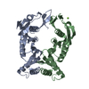
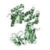
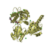
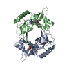
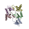
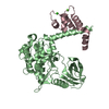
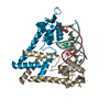
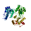
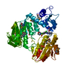
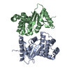
| Title | Crystal structure of the S210C (dimer) mutant from the N-terminal domain of the secretin XcpQ from Pseudomonas aeruginosa | ||||||
 Components Components | General secretion pathway protein D | ||||||
 Keywords Keywords |  PROTEIN TRANSPORT / XcpQ / structural protein / N-terminal domain of the secretin / periplasmic space / outer membrane PROTEIN TRANSPORT / XcpQ / structural protein / N-terminal domain of the secretin / periplasmic space / outer membrane | ||||||
| Function / homology |  Function and homology informationprotein secretion by the type II secretion system / type II protein secretion system complex / protein secretion / cell outer membrane / identical protein binding Function and homology informationprotein secretion by the type II secretion system / type II protein secretion system complex / protein secretion / cell outer membrane / identical protein bindingSimilarity search - Function | ||||||
| Biological species |   Pseudomonas aeruginosa (bacteria) Pseudomonas aeruginosa (bacteria) | ||||||
| Method | X-RAY DIFFRACTION / SYNCHROTRON / MOLECULAR REPLACEMENT / Resolution: 2.197 Å | ||||||
 Authors Authors | Van der Meeren, R. / Savvides, S. | ||||||
 Citation Citation | Journal: J.Biol.Chem. / Year: 2013 Title: New Insights into the Assembly of Bacterial Secretins: STRUCTURAL STUDIES OF THE PERIPLASMIC DOMAIN OF XcpQ FROM PSEUDOMONAS AERUGINOSA. Authors: Van der Meeren, R. / Wen, Y. / Van Gelder, P. / Tommassen, J. / Devreese, B. / Savvides, S.N. | ||||||
| History |
|
- Structure visualization
| Structure viewer | Molecule: MolmilJmol/JSmol |
|---|
- Downloads & links
-Download
| PDBx/mmCIF format | 4ec5.cif.gz | 152.2 KB | Display | PDBx/mmCIF format |
|---|---|---|---|---|
| PDB format | pdb4ec5.ent.gz | 127.9 KB | Display | PDB format |
| PDBx/mmJSON format | 4ec5.json.gz | Tree view | PDBx/mmJSON format | |
| Others |  Other downloads Other downloads |
-Validation report
| Arichive directory | https://data.pdbj.org/pub/pdb/validation_reports/ec/4ec5ftp://data.pdbj.org/pub/pdb/validation_reports/ec/4ec5 | HTTPS FTP |
|---|
-Related structure data

-Links
 PDBj
PDBj
- Assembly
| Deposited unit | 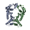
| ||||||||
|---|---|---|---|---|---|---|---|---|---|
| 1 |
| ||||||||
| 2 | 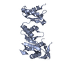
| ||||||||
| 3 | 
| ||||||||
| Unit cell |
| ||||||||
| Components on special symmetry positions |
|
-Components
| #1: Protein | Mass: 26535.959 Da / Num. of mol.: 2 / Fragment: UNP RESIDUES 35-277 / Mutation: S210C Source method: isolated from a genetically manipulated source Source: (gene. exp.) Pseudomonas aeruginosa (bacteria) / Strain: ATCC 15692/PAO1/1C/PRS 101/LMG 12228 / Gene: xcpQ, PA3105 / Production host: Escherichia coli (E. coli) / References: UniProt: P35818#2: Chemical | ChemComp-CA / |   Mass: 40.078 Da / Num. of mol.: 1 / Source method: obtained synthetically / Formula: Ca Mass: 40.078 Da / Num. of mol.: 1 / Source method: obtained synthetically / Formula: Ca#3: Water | ChemComp-HOH / | Water Mass: 18.015 Da / Num. of mol.: 141 / Source method: isolated from a natural source / Formula: H2O Mass: 18.015 Da / Num. of mol.: 141 / Source method: isolated from a natural source / Formula: H2O |
|---|
-Experimental details
-Experiment
| Experiment | Method: X-RAY DIFFRACTION / Number of used crystals: 1 |
|---|
- Sample preparation
| Crystal | Density Matthews: 2.09 Å3/Da / Density % sol: 41.28 % |
|---|---|
| Crystal grow | Temperature: 293.15 K / Method: vapor diffusion, sitting drop / pH: 7.5 Details: 0.1M HEPES pH 7.5, 14% PEG8000, 0.2M Calcium acetate hydrate, VAPOR DIFFUSION, SITTING DROP, temperature 293.15K |
-Data collection
| Diffraction source | Source: SYNCHROTRON / Site: ESRF  / Beamline: ID23-1 / Wavelength: 0.8726 Å / Beamline: ID23-1 / Wavelength: 0.8726 Å |
|---|---|
| Radiation | Protocol: SINGLE WAVELENGTH / Monochromatic (M) / Laue (L): M / Scattering type: x-ray |
| Radiation wavelength | Wavelength: 0.8726 Å / Relative weight: 1 |
| Reflection | Resolution: 2.197→50 Å / Num. all: 22844 / Num. obs: 22248 / % possible obs: 97.4 % / Observed criterion σ(F): 2 / Observed criterion σ(I): 2 |
- Processing
| Software |
| |||||||||||||||||||||||||||||||||||||||||||||||||||||||||||||||
|---|---|---|---|---|---|---|---|---|---|---|---|---|---|---|---|---|---|---|---|---|---|---|---|---|---|---|---|---|---|---|---|---|---|---|---|---|---|---|---|---|---|---|---|---|---|---|---|---|---|---|---|---|---|---|---|---|---|---|---|---|---|---|---|---|
| Refinement | Method to determine structure: MOLECULAR REPLACEMENT / Resolution: 2.197→46.602 Å / SU ML: 0.25 / σ(F): 1.99 / Phase error: 23.46 / Stereochemistry target values: ML
| |||||||||||||||||||||||||||||||||||||||||||||||||||||||||||||||
| Solvent computation | Shrinkage radii: 0.86 Å / VDW probe radii: 1.1 Å / Solvent model: FLAT BULK SOLVENT MODEL / Bsol: 27.277 Å2 / ksol: 0.324 e/Å3 | |||||||||||||||||||||||||||||||||||||||||||||||||||||||||||||||
| Displacement parameters |
| |||||||||||||||||||||||||||||||||||||||||||||||||||||||||||||||
| Refinement step | Cycle: LAST / Resolution: 2.197→46.602 Å
| |||||||||||||||||||||||||||||||||||||||||||||||||||||||||||||||
| Refine LS restraints |
| |||||||||||||||||||||||||||||||||||||||||||||||||||||||||||||||
| LS refinement shell |
|
