 Movie
Movie Controller


+ Open data

- Basic information
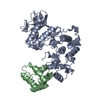
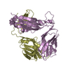
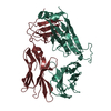
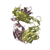
| Entry | Database: PDB / ID: 6paw | ||||||
|---|---|---|---|---|---|---|---|
| Title | Crystal structure of DAPK2 S308A Calcium/Calmodulin complex | ||||||
 Components Components |
| ||||||
 Keywords Keywords |  TRANSFERASE / APOPTOSIS / Kinase / Calcium signaling / Autophagy / Homodimer / Complex TRANSFERASE / APOPTOSIS / Kinase / Calcium signaling / Autophagy / Homodimer / Complex | ||||||
| Function / homology |  Function and homology information Function and homology informationautophagosome lumen / regulation of intrinsic apoptotic signaling pathway / positive regulation of eosinophil chemotaxis / Caspase activation via Dependence Receptors in the absence of ligand / positive regulation of neutrophil chemotaxis / anoikis / CaM pathway / Cam-PDE 1 activation / Sodium/Calcium exchangers / Calmodulin induced events ...autophagosome lumen / regulation of intrinsic apoptotic signaling pathway / positive regulation of eosinophil chemotaxis / Caspase activation via Dependence Receptors in the absence of ligand / positive regulation of neutrophil chemotaxis / anoikis / CaM pathway / Cam-PDE 1 activation / Sodium/Calcium exchangers / Calmodulin induced events / Reduction of cytosolic Ca++ levels / CREB1 phosphorylation through the activation of CaMKII/CaMKK/CaMKIV cascasde / Activation of Ca-permeable Kainate Receptor / Loss of phosphorylation of MECP2 at T308 / CREB1 phosphorylation through the activation of Adenylate Cyclase / PKA activation / negative regulation of high voltage-gated calcium channel activity / CaMK IV-mediated phosphorylation of CREB / Glycogen breakdown (glycogenolysis) / organelle localization by membrane tethering / negative regulation of calcium ion export across plasma membrane / Activation of RAC1 downstream of NMDARs / mitochondrion-endoplasmic reticulum membrane tethering / CLEC7A (Dectin-1) induces NFAT activation / regulation of cardiac muscle cell action potential / autophagosome membrane docking / positive regulation of ryanodine-sensitive calcium-release channel activity / regulation of cell communication by electrical coupling involved in cardiac conduction / Negative regulation of NMDA receptor-mediated neuronal transmission / negative regulation of peptidyl-threonine phosphorylation / Synthesis of IP3 and IP4 in the cytosol / Unblocking of NMDA receptors, glutamate binding and activation / Phase 0 - rapid depolarisation / protein phosphatase activator activity / RHO GTPases activate PAKs / positive regulation of cyclic-nucleotide phosphodiesterase activity / positive regulation of phosphoprotein phosphatase activity / Ion transport by P-type ATPases / Long-term potentiation / Uptake and function of anthrax toxins / Regulation of MECP2 expression and activity / Calcineurin activates NFAT / catalytic complex / DARPP-32 events / detection of calcium ion / negative regulation of ryanodine-sensitive calcium-release channel activity / Smooth Muscle Contraction / RHO GTPases activate IQGAPs / regulation of cardiac muscle contraction / calcium channel inhibitor activity / cellular response to interferon-beta / regulation of cardiac muscle contraction by regulation of the release of sequestered calcium ion / Protein methylation / eNOS activation / Activation of AMPK downstream of NMDARs / regulation of release of sequestered calcium ion into cytosol by sarcoplasmic reticulum / regulation of calcium-mediated signaling / Tetrahydrobiopterin (BH4) synthesis, recycling, salvage and regulation / positive regulation of protein dephosphorylation / voltage-gated potassium channel complex / Ion homeostasis / regulation of ryanodine-sensitive calcium-release channel activity / titin binding / positive regulation of protein autophosphorylation / sperm midpiece / calcium channel complex / substantia nigra development / adenylate cyclase activator activity / Ras activation upon Ca2+ influx through NMDA receptor / regulation of heart rate / sarcomere / protein serine/threonine kinase activator activity / FCERI mediated Ca+2 mobilization / FCGR3A-mediated IL10 synthesis / VEGFR2 mediated vascular permeability / positive regulation of peptidyl-threonine phosphorylation / regulation of cytokinesis / Antigen activates B Cell Receptor (BCR) leading to generation of second messengers / VEGFR2 mediated cell proliferation / regulation of autophagy / Translocation of SLC2A4 (GLUT4) to the plasma membrane / RAF activation / positive regulation of receptor signaling pathway via JAK-STAT / Transcriptional activation of mitochondrial biogenesis / positive regulation of protein serine/threonine kinase activity / spindle microtubule / Stimuli-sensing channels / cellular response to type II interferon / spindle pole / response to calcium ion / RAS processing / Signaling by RAF1 mutants / Signaling by moderate kinase activity BRAF mutants / Paradoxical activation of RAF signaling by kinase inactive BRAF / Signaling downstream of RAS mutants / Inactivation, recovery and regulation of the phototransduction cascade / calcium-dependent protein binding / G2/M transition of mitotic cell cycle / Signaling by BRAF and RAF1 fusions / Platelet degranulation Similarity search - Function | ||||||
| Biological species |  Homo sapiens (human) Homo sapiens (human) | ||||||
| Method | X-RAY DIFFRACTION / SYNCHROTRON / MOLECULAR REPLACEMENT / Resolution: 2.953 Å | ||||||
 Authors Authors | Simon, B. / Wilmanns, M. | ||||||
 Citation Citation | Journal: To Be Published Title: Crystal structure of Death-associated protein kinase 2 in complex with Calcium Calmodulin Authors: Simon, B. / Wilmanns, M. | ||||||
| History |
|
- Structure visualization
| Structure viewer | Molecule: MolmilJmol/JSmol |
|---|
- Downloads & links
-Download
| PDBx/mmCIF format | 6paw.cif.gz | 330.8 KB | Display | PDBx/mmCIF format |
|---|---|---|---|---|
| PDB format | pdb6paw.ent.gz | 265.5 KB | Display | PDB format |
| PDBx/mmJSON format | 6paw.json.gz | Tree view | PDBx/mmJSON format | |
| Others |  Other downloads Other downloads |
-Validation report
| Arichive directory | https://data.pdbj.org/pub/pdb/validation_reports/pa/6pawftp://data.pdbj.org/pub/pdb/validation_reports/pa/6paw | HTTPS FTP |
|---|
-Related structure data


-Links
 PDBj
PDBj


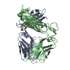
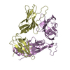
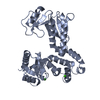
- Assembly
| Deposited unit | 
| ||||||||
|---|---|---|---|---|---|---|---|---|---|
| 1 | 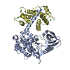
| ||||||||
| 2 | 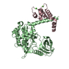
| ||||||||
| 3 | 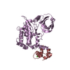
| ||||||||
| 4 | 
| ||||||||
| Unit cell |
|
-Components
| #1: Protein | Mass: 37375.727 Da / Num. of mol.: 4 / Mutation: S308A Source method: isolated from a genetically manipulated source Source: (gene. exp.) Homo sapiens (human) / Gene: DAPK2 / Plasmid: pnEK / Production host:  Escherichia coli BL21 (bacteria) Escherichia coli BL21 (bacteria)References: UniProt: Q9UIK4, non-specific serine/threonine protein kinase#2: Protein | Mass: 16852.545 Da / Num. of mol.: 4 Source method: isolated from a genetically manipulated source Source: (gene. exp.) Homo sapiens (human) / Gene: CALM1, CALM, CAM, CAM1 / Plasmid: pnEK / Production host: Escherichia coli BL21 (bacteria) / References: UniProt: P0DP23#3: Chemical | ChemComp-CA /   Mass: 40.078 Da / Num. of mol.: 9 / Source method: obtained synthetically / Formula: Ca Mass: 40.078 Da / Num. of mol.: 9 / Source method: obtained synthetically / Formula: CaHas ligand of interest | N | |
|---|
-Experimental details
-Experiment
| Experiment | Method: X-RAY DIFFRACTION / Number of used crystals: 1 |
|---|
- Sample preparation
| Crystal | Density Matthews: 2.6 Å3/Da / Density % sol: 52.73 % |
|---|---|
| Crystal grow | Temperature: 292.15 K / Method: vapor diffusion, sitting drop / pH: 6.5 Details: 0.1 M Bis-Tris-HCl, pH 6.5, 0.6 M sodium chloride, 14.3% PEG3350 |
-Data collection
| Diffraction | Mean temperature: 100 K / Serial crystal experiment: N |
|---|---|
| Diffraction source | Source: SYNCHROTRON / Site: PETRA III, EMBL c/o DESY  / Beamline: P14 (MX2) / Wavelength: 0.9763 Å / Beamline: P14 (MX2) / Wavelength: 0.9763 Å |
| Detector | Type: DECTRIS EIGER X 16M / Detector: PIXEL / Date: Jun 6, 2015 |
| Radiation | Protocol: SINGLE WAVELENGTH / Monochromatic (M) / Laue (L): M / Scattering type: x-ray |
| Radiation wavelength | Wavelength: 0.9763 Å / Relative weight: 1 |
| Reflection | Resolution: 2.95→131.65 Å / Num. obs: 48427 / % possible obs: 64.4 % / Redundancy: 8.4 % / Rmerge(I) obs: 0.275 / Net I/σ(I): 9.5 |
| Reflection shell | Resolution: 2.95→3.2 Å / Rmerge(I) obs: 1.853 / Mean I/σ(I) obs: 1.4 / Num. unique obs: 1250 |
- Processing
| Software |
| ||||||||||||||||||||||||||||||||||||||||||||||||||||||||||||||||||||||||||||||||||||
|---|---|---|---|---|---|---|---|---|---|---|---|---|---|---|---|---|---|---|---|---|---|---|---|---|---|---|---|---|---|---|---|---|---|---|---|---|---|---|---|---|---|---|---|---|---|---|---|---|---|---|---|---|---|---|---|---|---|---|---|---|---|---|---|---|---|---|---|---|---|---|---|---|---|---|---|---|---|---|---|---|---|---|---|---|---|
| Refinement | Method to determine structure: MOLECULAR REPLACEMENT Starting model: PDB entries 2A2A & 1ZUZ Resolution: 2.953→131.65 Å / SU ML: 0.36 / Cross valid method: THROUGHOUT / σ(F): 1.36 / Phase error: 31.61
| ||||||||||||||||||||||||||||||||||||||||||||||||||||||||||||||||||||||||||||||||||||
| Solvent computation | Shrinkage radii: 0.9 Å / VDW probe radii: 1.11 Å | ||||||||||||||||||||||||||||||||||||||||||||||||||||||||||||||||||||||||||||||||||||
| Displacement parameters | Biso max: 159.8 Å2 / Biso mean: 57.7375 Å2 / Biso min: 12.11 Å2 | ||||||||||||||||||||||||||||||||||||||||||||||||||||||||||||||||||||||||||||||||||||
| Refinement step | Cycle: final / Resolution: 2.953→131.65 Å
| ||||||||||||||||||||||||||||||||||||||||||||||||||||||||||||||||||||||||||||||||||||
| LS refinement shell | Refine-ID: X-RAY DIFFRACTION / Rfactor Rfree error: 0 / Total num. of bins used: 11
|
