 Movie
Movie Controller

[English] 日本語
 Yorodumi
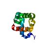
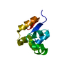
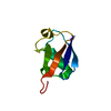
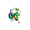
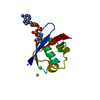
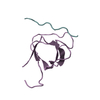
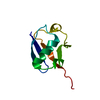
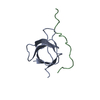
- PDB-4dwf: Crystal structure of a HLA-B associated transcript 3 (BAT3) from ... -
+ Open data

- Basic information
| Entry | Database: PDB / ID: 4dwf | ||||||
|---|---|---|---|---|---|---|---|
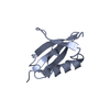
| Title | Crystal structure of a HLA-B associated transcript 3 (BAT3) from Homo sapiens at 1.80 A resolution | ||||||
 Components Components | HLA-B-associated transcript 3 | ||||||
 Keywords Keywords |  STRUCTURAL GENOMICS / UNKNOWN FUNCTION / Ubiquitin-like domain / BAT3 protein / PF00240 / Joint Center for Structural Genomics / JCSG / Protein Structure Initiative / PSI-BIOLOGY / Partnership for T-Cell Biology / TCELL STRUCTURAL GENOMICS / UNKNOWN FUNCTION / Ubiquitin-like domain / BAT3 protein / PF00240 / Joint Center for Structural Genomics / JCSG / Protein Structure Initiative / PSI-BIOLOGY / Partnership for T-Cell Biology / TCELL | ||||||
| Function / homology |  Function and homology informationBAT3 complex / immune response-activating cell surface receptor signaling pathway / maintenance of unfolded protein / : / positive regulation of ERAD pathway / tail-anchored membrane protein insertion into ER membrane / synaptonemal complex assembly / post-translational protein targeting to endoplasmic reticulum membrane / internal peptidyl-lysine acetylation / misfolded protein binding ...BAT3 complex / immune response-activating cell surface receptor signaling pathway / maintenance of unfolded protein / : / positive regulation of ERAD pathway / tail-anchored membrane protein insertion into ER membrane / synaptonemal complex assembly / post-translational protein targeting to endoplasmic reticulum membrane / internal peptidyl-lysine acetylation / misfolded protein binding / natural killer cell activation / endoplasmic reticulum stress-induced pre-emptive quality control / ERAD pathway / Insertion of tail-anchored proteins into the endoplasmic reticulum membrane / proteasome binding / ubiquitin-specific protease binding / : / regulation of embryonic development / negative regulation of proteasomal ubiquitin-dependent protein catabolic process / polyubiquitin modification-dependent protein binding / intrinsic apoptotic signaling pathway in response to endoplasmic reticulum stress / intrinsic apoptotic signaling pathway in response to DNA damage by p53 class mediator / Hsp70 protein binding / kidney development / proteasomal protein catabolic process / negative regulation of proteolysis / lung development / regulation of protein stability / brain development / ribosome binding / chromatin organization / ubiquitin-dependent protein catabolic process / spermatogenesis / cell differentiation / protein stabilization / signaling receptor binding / intracellular membrane-bounded organelle / apoptotic process / ubiquitin protein ligase binding / negative regulation of apoptotic process / extracellular exosome / nucleoplasm / membrane / identical protein binding / nucleus / cytosol / cytoplasm Function and homology informationBAT3 complex / immune response-activating cell surface receptor signaling pathway / maintenance of unfolded protein / : / positive regulation of ERAD pathway / tail-anchored membrane protein insertion into ER membrane / synaptonemal complex assembly / post-translational protein targeting to endoplasmic reticulum membrane / internal peptidyl-lysine acetylation / misfolded protein binding ...BAT3 complex / immune response-activating cell surface receptor signaling pathway / maintenance of unfolded protein / : / positive regulation of ERAD pathway / tail-anchored membrane protein insertion into ER membrane / synaptonemal complex assembly / post-translational protein targeting to endoplasmic reticulum membrane / internal peptidyl-lysine acetylation / misfolded protein binding / natural killer cell activation / endoplasmic reticulum stress-induced pre-emptive quality control / ERAD pathway / Insertion of tail-anchored proteins into the endoplasmic reticulum membrane / proteasome binding / ubiquitin-specific protease binding / : / regulation of embryonic development / negative regulation of proteasomal ubiquitin-dependent protein catabolic process / polyubiquitin modification-dependent protein binding / intrinsic apoptotic signaling pathway in response to endoplasmic reticulum stress / intrinsic apoptotic signaling pathway in response to DNA damage by p53 class mediator / Hsp70 protein binding / kidney development / proteasomal protein catabolic process / negative regulation of proteolysis / lung development / regulation of protein stability / brain development / ribosome binding / chromatin organization / ubiquitin-dependent protein catabolic process / spermatogenesis / cell differentiation / protein stabilization / signaling receptor binding / intracellular membrane-bounded organelle / apoptotic process / ubiquitin protein ligase binding / negative regulation of apoptotic process / extracellular exosome / nucleoplasm / membrane / identical protein binding / nucleus / cytosol / cytoplasmSimilarity search - Function | ||||||
| Biological species |  Homo sapiens (human) Homo sapiens (human) | ||||||
| Method | X-RAY DIFFRACTION / SYNCHROTRON / MAD / Resolution: 1.8 Å | ||||||
 Authors Authors | Joint Center for Structural Genomics (JCSG) / Partnership for T-Cell Biology (TCELL) | ||||||
 Citation Citation | Journal: To be published Title: Crystal structure of a HLA-B associated transcript 3 (BAT3) from Homo sapiens at 1.80 A resolution Authors: Joint Center for Structural Genomics (JCSG) / Partnership for T-Cell Biology (TCELL) | ||||||
| History |
|
- Structure visualization
| Structure viewer | Molecule: MolmilJmol/JSmol |
|---|
- Downloads & links
-Download
| PDBx/mmCIF format | 4dwf.cif.gz | 78.7 KB | Display | PDBx/mmCIF format |
|---|---|---|---|---|
| PDB format | pdb4dwf.ent.gz | 61.8 KB | Display | PDB format |
| PDBx/mmJSON format | 4dwf.json.gz | Tree view | PDBx/mmJSON format | |
| Others |  Other downloads Other downloads |
-Validation report
| Arichive directory | https://data.pdbj.org/pub/pdb/validation_reports/dw/4dwfftp://data.pdbj.org/pub/pdb/validation_reports/dw/4dwf | HTTPS FTP |
|---|
-Related structure data
| Similar structure data | |
|---|---|
| Other databases |
-Links
 PDBj
PDBj


- Assembly
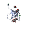
| Deposited unit | 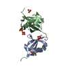
| ||||||||
|---|---|---|---|---|---|---|---|---|---|
| 1 | 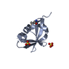
| ||||||||
| 2 | 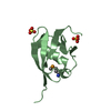
| ||||||||
| Unit cell |
| ||||||||
| Details | CRYSTAL PACKING ANALYSIS SUGGESTS THE ASSIGNMENT OF A MONOMER AS THE SIGNIFICANT OLIGOMERIZATION STATE. |
-Components
| #1: Protein | Mass: 9958.121 Da / Num. of mol.: 2 Source method: isolated from a genetically manipulated source Source: (gene. exp.) Homo sapiens (human) / Gene: BAG6, BAT3, BC003133, G3 / Plasmid: SpeedET / Production host:  Escherichia Coli (E. coli) / Strain (production host): HK100 / References: UniProt: P46379 Escherichia Coli (E. coli) / Strain (production host): HK100 / References: UniProt: P46379#2: Chemical | ChemComp-SO4 / Sulfate  Mass: 96.063 Da / Num. of mol.: 4 / Source method: obtained synthetically / Formula: SO4 Mass: 96.063 Da / Num. of mol.: 4 / Source method: obtained synthetically / Formula: SO4#3: Water | ChemComp-HOH / | Water Mass: 18.015 Da / Num. of mol.: 170 / Source method: isolated from a natural source / Formula: H2O Mass: 18.015 Da / Num. of mol.: 170 / Source method: isolated from a natural source / Formula: H2OSequence details | THIS CONSTRUCT WAS EXPRESSED WITH A PURIFICATION TAG MGSDKIHHHHHHENLYFQG. THE TAG WAS REMOVED WITH ...THIS CONSTRUCT WAS EXPRESSED WITH A PURIFICATI | |
|---|
-Experimental details
-Experiment
| Experiment | Method: X-RAY DIFFRACTION / Number of used crystals: 1 |
|---|
- Sample preparation
| Crystal | Density Matthews: 2.46 Å3/Da / Density % sol: 49.98 % |
|---|---|
| Crystal grow | Temperature: 277 K / Method: vapor diffusion, sitting drop / pH: 5.6 Details: 2.0M (NH4)2SO4, 0.2M K/Na Tartrate, 0.1M Citrate pH 5.6, NANODROP, VAPOR DIFFUSION, SITTING DROP, temperature 277K |
-Data collection
| Diffraction | Mean temperature: 100 K | |||||||||||||||||||||||||||||||||||||||||||||||||||||||||||||||||||||||||||||
|---|---|---|---|---|---|---|---|---|---|---|---|---|---|---|---|---|---|---|---|---|---|---|---|---|---|---|---|---|---|---|---|---|---|---|---|---|---|---|---|---|---|---|---|---|---|---|---|---|---|---|---|---|---|---|---|---|---|---|---|---|---|---|---|---|---|---|---|---|---|---|---|---|---|---|---|---|---|---|
| Diffraction source | Source: SYNCHROTRON / Site: SSRL  / Beamline: BL9-2 / Wavelength: 0.91162, 0.97949, 0.97903 / Beamline: BL9-2 / Wavelength: 0.91162, 0.97949, 0.97903 | |||||||||||||||||||||||||||||||||||||||||||||||||||||||||||||||||||||||||||||
| Detector | Type: MARMOSAIC 325 mm CCD / Detector: CCD / Date: Dec 1, 2011 / Details: double crystal monochromator | |||||||||||||||||||||||||||||||||||||||||||||||||||||||||||||||||||||||||||||
| Radiation | Monochromator: double crystal / Protocol: MAD / Monochromatic (M) / Laue (L): M / Scattering type: x-ray | |||||||||||||||||||||||||||||||||||||||||||||||||||||||||||||||||||||||||||||
| Radiation wavelength |
| |||||||||||||||||||||||||||||||||||||||||||||||||||||||||||||||||||||||||||||
| Reflection | Resolution: 1.8→28.33 Å / Num. obs: 18720 / % possible obs: 97.9 % / Observed criterion σ(I): -3 / Biso Wilson estimate: 26.113 Å2 / Rmerge(I) obs: 0.042 / Net I/σ(I): 10.74 | |||||||||||||||||||||||||||||||||||||||||||||||||||||||||||||||||||||||||||||
| Reflection shell | Diffraction-ID: 1
|
-Phasing
| Phasing | Method: MAD |
|---|
- Processing
| Software |
| |||||||||||||||||||||||||||||||||||||||||||||||||||||||||||||||||||||||||||
|---|---|---|---|---|---|---|---|---|---|---|---|---|---|---|---|---|---|---|---|---|---|---|---|---|---|---|---|---|---|---|---|---|---|---|---|---|---|---|---|---|---|---|---|---|---|---|---|---|---|---|---|---|---|---|---|---|---|---|---|---|---|---|---|---|---|---|---|---|---|---|---|---|---|---|---|---|
| Refinement | Method to determine structure: MAD / Resolution: 1.8→28.33 Å / Cor.coef. Fo:Fc: 0.972 / Cor.coef. Fo:Fc free: 0.946 / Occupancy max: 1 / Occupancy min: 0.4 / SU B: 6.014 / SU ML: 0.094 / Cross valid method: THROUGHOUT / σ(F): 0 / ESU R: 0.118 / ESU R Free: 0.127 Stereochemistry target values: MAXIMUM LIKELIHOOD WITH PHASES Details: 1. HYDROGENS HAVE BEEN ADDED IN THE RIDING POSITIONS. 2. ATOM RECORD CONTAINS SUM OF TLS AND RESIDUAL B FACTORS. 3. ANISOU RECORD CONTAINS SUM OF TLS AND RESIDUAL U FACTORS. 4. WATERS WERE ...Details: 1. HYDROGENS HAVE BEEN ADDED IN THE RIDING POSITIONS. 2. ATOM RECORD CONTAINS SUM OF TLS AND RESIDUAL B FACTORS. 3. ANISOU RECORD CONTAINS SUM OF TLS AND RESIDUAL U FACTORS. 4. WATERS WERE EXCLUDED FROM AUTOMATIC TLS ASSIGNMENT. 5. A MET-INHIBITION PROTOCOL WAS USED FOR SELENOMETHIONINE INCORPORATION DURING PROTEIN EXPRESSION. THE OCCUPANCY OF THE SE ATOMS IN THE MSE RESIDUES WAS REDUCED TO 0.75 FOR THE REDUCED SCATTERING POWER DUE TO PARTIAL S-MET INCORPORATION. 6. SULFATE IONS (SO4) FROM THE CRYSTALLIZATION SOLUTION ARE MODELED.
| |||||||||||||||||||||||||||||||||||||||||||||||||||||||||||||||||||||||||||
| Solvent computation | Ion probe radii: 0.8 Å / Shrinkage radii: 0.8 Å / VDW probe radii: 1.2 Å / Solvent model: BABINET MODEL WITH MASK | |||||||||||||||||||||||||||||||||||||||||||||||||||||||||||||||||||||||||||
| Displacement parameters | Biso max: 99 Å2 / Biso mean: 37.9515 Å2 / Biso min: 21.75 Å2
| |||||||||||||||||||||||||||||||||||||||||||||||||||||||||||||||||||||||||||
| Refinement step | Cycle: LAST / Resolution: 1.8→28.33 Å
| |||||||||||||||||||||||||||||||||||||||||||||||||||||||||||||||||||||||||||
| Refine LS restraints |
| |||||||||||||||||||||||||||||||||||||||||||||||||||||||||||||||||||||||||||
| LS refinement shell | Resolution: 1.8→1.847 Å / Total num. of bins used: 20
| |||||||||||||||||||||||||||||||||||||||||||||||||||||||||||||||||||||||||||
| Refinement TLS params. | Method: refined / Refine-ID: X-RAY DIFFRACTION
| |||||||||||||||||||||||||||||||||||||||||||||||||||||||||||||||||||||||||||
| Refinement TLS group |
|
