 Movie
Movie Controller

[English] 日本語
 Yorodumi
- PDB-4dhl: Crystal structure of red kidney bean purple acid phosphatase in c... -
+ Open data

- Basic information
| Entry | Database: PDB / ID: 4dhl | |||||||||
|---|---|---|---|---|---|---|---|---|---|---|
| Title | Crystal structure of red kidney bean purple acid phosphatase in complex with Maybridge fragment MO07123 | |||||||||
 Components Components | Purple acid phosphatase | |||||||||
 Keywords Keywords |  HYDROLASE / Catalytic C domain / N terminal domain / Phosphatase / Acid Phosphatase / Phosphoric Monoester Hydrolases / MO07123 / Maybridge / Fragment / lysosome HYDROLASE / Catalytic C domain / N terminal domain / Phosphatase / Acid Phosphatase / Phosphoric Monoester Hydrolases / MO07123 / Maybridge / Fragment / lysosome | |||||||||
| Function / homology |  Function and homology informationacid phosphatase / acid phosphatase activity / ferric iron binding / zinc ion binding / extracellular region Function and homology informationacid phosphatase / acid phosphatase activity / ferric iron binding / zinc ion binding / extracellular regionSimilarity search - Function | |||||||||
| Biological species |  Phaseolus vulgaris (French bean) Phaseolus vulgaris (French bean) | |||||||||
| Method | X-RAY DIFFRACTION / SYNCHROTRON / MOLECULAR REPLACEMENT / Resolution: 2.3 Å | |||||||||
 Authors Authors | Feder, D. / Clayton, D.J. / Hussein, W.M. / Schenk, G. / McGeary, R. / Guddat, L.W. | |||||||||
 Citation Citation | Journal: Chem.Biol.Drug Des. / Year: 2012 Title: Identification of purple acid phosphatase inhibitors by fragment-based screening: promising new leads for osteoporosis therapeutics. Authors: Feder, D. / Hussein, W.M. / Clayton, D.J. / Kan, M.W. / Schenk, G. / McGeary, R.P. / Guddat, L.W. | |||||||||
| History |
|
- Structure visualization
| Structure viewer | Molecule: MolmilJmol/JSmol |
|---|
- Downloads & links
-Download
| PDBx/mmCIF format | 4dhl.cif.gz | 394.6 KB | Display | PDBx/mmCIF format |
|---|---|---|---|---|
| PDB format | pdb4dhl.ent.gz | 326.6 KB | Display | PDB format |
| PDBx/mmJSON format | 4dhl.json.gz | Tree view | PDBx/mmJSON format | |
| Others |  Other downloads Other downloads |
-Validation report
| Arichive directory | https://data.pdbj.org/pub/pdb/validation_reports/dh/4dhlftp://data.pdbj.org/pub/pdb/validation_reports/dh/4dhl | HTTPS FTP |
|---|
-Related structure data


-Links
 PDBj
PDBj
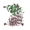
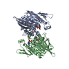
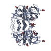
- Assembly
| Deposited unit | 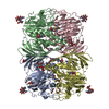
| ||||||||
|---|---|---|---|---|---|---|---|---|---|
| 1 | 
| ||||||||
| 2 | 
| ||||||||
| Unit cell |
|
-Components
-Protein , 1 types, 4 molecules ABDC
| #1: Protein | Mass: 49555.398 Da / Num. of mol.: 4 / Source method: isolated from a natural source / Source: (natural) Phaseolus vulgaris (French bean)References: UniProt: O24319, UniProt: P80366*PLUS, acid phosphatase |
|---|
-Sugars , 3 types, 16 molecules 
| #2: Polysaccharide | alpha-L-fucopyranose-(1-3)-[2-acetamido-2-deoxy-beta-D-glucopyranose-(1-4)]2-acetamido-2-deoxy-beta- ...alpha-L-fucopyranose-(1-3)-[2-acetamido-2-deoxy-beta-D-glucopyranose-(1-4)]2-acetamido-2-deoxy-beta-D-glucopyranose / Mass: 570.542 Da / Num. of mol.: 6Source method: isolated from a genetically manipulated source #3: Polysaccharide | / Mass: 367.349 Da / Num. of mol.: 3Source method: isolated from a genetically manipulated source #10: Sugar | ChemComp-NAG / N-Acetylglucosamine Type: D-saccharide, beta linking / Mass: 221.208 Da / Num. of mol.: 7 Type: D-saccharide, beta linking / Mass: 221.208 Da / Num. of mol.: 7Source method: isolated from a genetically manipulated source Formula: C8H15NO6 |
|---|
-Non-polymers , 7 types, 1571 molecules 


| #4: Chemical | ChemComp-ZN /  Mass: 65.409 Da / Num. of mol.: 4 / Source method: obtained synthetically / Formula: Zn Mass: 65.409 Da / Num. of mol.: 4 / Source method: obtained synthetically / Formula: Zn#5: Chemical | ChemComp-FE / Iron Mass: 55.845 Da / Num. of mol.: 4 / Source method: obtained synthetically / Formula: Fe Mass: 55.845 Da / Num. of mol.: 4 / Source method: obtained synthetically / Formula: Fe#6: Chemical | ChemComp-0K7 /  Mass: 219.260 Da / Num. of mol.: 4 / Source method: obtained synthetically / Formula: C11H9NO2S Mass: 219.260 Da / Num. of mol.: 4 / Source method: obtained synthetically / Formula: C11H9NO2S#7: Chemical | Glycerol Mass: 92.094 Da / Num. of mol.: 3 / Source method: obtained synthetically / Formula: C3H8O3 Mass: 92.094 Da / Num. of mol.: 3 / Source method: obtained synthetically / Formula: C3H8O3#8: Chemical | ChemComp-SO4 / Sulfate Mass: 96.063 Da / Num. of mol.: 5 / Source method: obtained synthetically / Formula: SO4 Mass: 96.063 Da / Num. of mol.: 5 / Source method: obtained synthetically / Formula: SO4#9: Chemical | ChemComp-EDO / Ethylene glycol Mass: 62.068 Da / Num. of mol.: 6 / Source method: obtained synthetically / Formula: C2H6O2 Mass: 62.068 Da / Num. of mol.: 6 / Source method: obtained synthetically / Formula: C2H6O2#11: Water | ChemComp-HOH / | WaterMass: 18.015 Da / Num. of mol.: 1545 / Source method: isolated from a natural source / Formula: H2O |
|---|
-Experimental details
-Experiment
| Experiment | Method: X-RAY DIFFRACTION / Number of used crystals: 1 |
|---|
- Sample preparation
| Crystal | Density Matthews: 3.45 Å3/Da / Density % sol: 64.35 % |
|---|---|
| Crystal grow | Temperature: 291 K / Method: vapor diffusion, hanging drop / pH: 4 Details: 2.3M ammonium sulfate, 0.1M sodium acetate pH 4.0, VAPOR DIFFUSION, HANGING DROP, temperature 291K |
-Data collection
| Diffraction source | Source: SYNCHROTRON / Site: Australian Synchrotron  / Beamline: MX2 / Wavelength: 0.9 Å / Beamline: MX2 / Wavelength: 0.9 Å |
|---|---|
| Radiation | Protocol: SINGLE WAVELENGTH / Monochromatic (M) / Laue (L): M / Scattering type: x-ray |
| Radiation wavelength | Wavelength: 0.9 Å / Relative weight: 1 |
| Reflection | Resolution: 2.3→19.82 Å / Num. all: 115253 / Num. obs: 115053 |
- Processing
| Software | Name: PHENIX / Version: (phenix.refine: 1.7_650) / Classification: refinement | |||||||||||||||||||||||||||||||||||||||||||||||||||||||||||||||||||||||||||||||||||||||||||||||||||||||||||||||||||||||||||||||||||||||||||||||||||||||||||||||||||||||||||||||||||||||||||||||||||||||||||||||||||||||||
|---|---|---|---|---|---|---|---|---|---|---|---|---|---|---|---|---|---|---|---|---|---|---|---|---|---|---|---|---|---|---|---|---|---|---|---|---|---|---|---|---|---|---|---|---|---|---|---|---|---|---|---|---|---|---|---|---|---|---|---|---|---|---|---|---|---|---|---|---|---|---|---|---|---|---|---|---|---|---|---|---|---|---|---|---|---|---|---|---|---|---|---|---|---|---|---|---|---|---|---|---|---|---|---|---|---|---|---|---|---|---|---|---|---|---|---|---|---|---|---|---|---|---|---|---|---|---|---|---|---|---|---|---|---|---|---|---|---|---|---|---|---|---|---|---|---|---|---|---|---|---|---|---|---|---|---|---|---|---|---|---|---|---|---|---|---|---|---|---|---|---|---|---|---|---|---|---|---|---|---|---|---|---|---|---|---|---|---|---|---|---|---|---|---|---|---|---|---|---|---|---|---|---|---|---|---|---|---|---|---|---|---|---|---|---|---|---|---|---|
| Refinement | Method to determine structure: MOLECULAR REPLACEMENT / Resolution: 2.3→19.82 Å / SU ML: 0.28 / σ(F): 0.34 / Phase error: 17.68 / Stereochemistry target values: ML
| |||||||||||||||||||||||||||||||||||||||||||||||||||||||||||||||||||||||||||||||||||||||||||||||||||||||||||||||||||||||||||||||||||||||||||||||||||||||||||||||||||||||||||||||||||||||||||||||||||||||||||||||||||||||||
| Solvent computation | Shrinkage radii: 0.9 Å / VDW probe radii: 1.11 Å / Solvent model: FLAT BULK SOLVENT MODEL / Bsol: 40.758 Å2 / ksol: 0.358 e/Å3 | |||||||||||||||||||||||||||||||||||||||||||||||||||||||||||||||||||||||||||||||||||||||||||||||||||||||||||||||||||||||||||||||||||||||||||||||||||||||||||||||||||||||||||||||||||||||||||||||||||||||||||||||||||||||||
| Displacement parameters |
| |||||||||||||||||||||||||||||||||||||||||||||||||||||||||||||||||||||||||||||||||||||||||||||||||||||||||||||||||||||||||||||||||||||||||||||||||||||||||||||||||||||||||||||||||||||||||||||||||||||||||||||||||||||||||
| Refinement step | Cycle: LAST / Resolution: 2.3→19.82 Å
| |||||||||||||||||||||||||||||||||||||||||||||||||||||||||||||||||||||||||||||||||||||||||||||||||||||||||||||||||||||||||||||||||||||||||||||||||||||||||||||||||||||||||||||||||||||||||||||||||||||||||||||||||||||||||
| Refine LS restraints |
| |||||||||||||||||||||||||||||||||||||||||||||||||||||||||||||||||||||||||||||||||||||||||||||||||||||||||||||||||||||||||||||||||||||||||||||||||||||||||||||||||||||||||||||||||||||||||||||||||||||||||||||||||||||||||
| LS refinement shell |
|
