 Movie
Movie Controller

[English] 日本語
 Yorodumi
- PDB-4dh0: X-ray Crystal Structure of 28-O-Methylrapamycin complexed with FK... -
+ Open data

- Basic information
| Entry | Database: PDB / ID: 4dh0 | ||||||
|---|---|---|---|---|---|---|---|
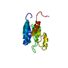
| Title | X-ray Crystal Structure of 28-O-Methylrapamycin complexed with FKBP12: Is the Cyclohexyl Moiety Part of the Effector Domain of Rapamycin? | ||||||
 Components Components | Peptidyl-prolyl cis-trans isomerase FKBP1A | ||||||
 Keywords Keywords | ISOMERASE/IMMUNOSUPPRESSANT /  peptidyl-prolyl cis-trans isomerase / ISOMERASE-IMMUNOSUPPRESSANT complex peptidyl-prolyl cis-trans isomerase / ISOMERASE-IMMUNOSUPPRESSANT complex | ||||||
| Function / homology |  Function and homology informationmacrolide binding / activin receptor binding / cytoplasmic side of membrane / signaling receptor inhibitor activity / transforming growth factor beta receptor binding / TGFBR1 LBD Mutants in Cancer / type I transforming growth factor beta receptor binding / negative regulation of activin receptor signaling pathway / heart trabecula formation / terminal cisterna ...macrolide binding / activin receptor binding / cytoplasmic side of membrane / signaling receptor inhibitor activity / transforming growth factor beta receptor binding / TGFBR1 LBD Mutants in Cancer / type I transforming growth factor beta receptor binding / negative regulation of activin receptor signaling pathway / heart trabecula formation / terminal cisterna / ryanodine receptor complex / I-SMAD binding / regulation of amyloid precursor protein catabolic process / protein maturation by protein folding / 'de novo' protein folding / ventricular cardiac muscle tissue morphogenesis / negative regulation of phosphoprotein phosphatase activity / FK506 binding / mTORC1-mediated signalling / TGF-beta receptor signaling activates SMADs / Calcineurin activates NFAT / regulation of immune response / protein peptidyl-prolyl isomerization / supramolecular fiber organization / heart morphogenesis / regulation of ryanodine-sensitive calcium-release channel activity / sarcoplasmic reticulum membrane / T cell activation / TGF-beta receptor signaling in EMT (epithelial to mesenchymal transition) / sarcoplasmic reticulum / peptidylprolyl isomerase / peptidyl-prolyl cis-trans isomerase activity / calcium ion transmembrane transport / negative regulation of transforming growth factor beta receptor signaling pathway / Z disc / SARS-CoV-1 activates/modulates innate immune responses / regulation of protein localization / protein folding / positive regulation of protein binding / protein refolding / positive regulation of canonical NF-kappaB signal transduction / transmembrane transporter binding / Potential therapeutics for SARS / amyloid fibril formation / membrane / cytosol / cytoplasm Function and homology informationmacrolide binding / activin receptor binding / cytoplasmic side of membrane / signaling receptor inhibitor activity / transforming growth factor beta receptor binding / TGFBR1 LBD Mutants in Cancer / type I transforming growth factor beta receptor binding / negative regulation of activin receptor signaling pathway / heart trabecula formation / terminal cisterna ...macrolide binding / activin receptor binding / cytoplasmic side of membrane / signaling receptor inhibitor activity / transforming growth factor beta receptor binding / TGFBR1 LBD Mutants in Cancer / type I transforming growth factor beta receptor binding / negative regulation of activin receptor signaling pathway / heart trabecula formation / terminal cisterna / ryanodine receptor complex / I-SMAD binding / regulation of amyloid precursor protein catabolic process / protein maturation by protein folding / 'de novo' protein folding / ventricular cardiac muscle tissue morphogenesis / negative regulation of phosphoprotein phosphatase activity / FK506 binding / mTORC1-mediated signalling / TGF-beta receptor signaling activates SMADs / Calcineurin activates NFAT / regulation of immune response / protein peptidyl-prolyl isomerization / supramolecular fiber organization / heart morphogenesis / regulation of ryanodine-sensitive calcium-release channel activity / sarcoplasmic reticulum membrane / T cell activation / TGF-beta receptor signaling in EMT (epithelial to mesenchymal transition) / sarcoplasmic reticulum / peptidylprolyl isomerase / peptidyl-prolyl cis-trans isomerase activity / calcium ion transmembrane transport / negative regulation of transforming growth factor beta receptor signaling pathway / Z disc / SARS-CoV-1 activates/modulates innate immune responses / regulation of protein localization / protein folding / positive regulation of protein binding / protein refolding / positive regulation of canonical NF-kappaB signal transduction / transmembrane transporter binding / Potential therapeutics for SARS / amyloid fibril formation / membrane / cytosol / cytoplasmSimilarity search - Function | ||||||
| Biological species |  Homo sapiens (human) Homo sapiens (human) | ||||||
| Method | X-RAY DIFFRACTION / FOURIER SYNTHESIS / Resolution: 2.1 Å | ||||||
 Authors Authors | Kallen, J. | ||||||
 Citation Citation | Journal: J.Am.Chem.Soc. / Year: 1996 Title: X-ray Crystal Structure of 28-O-Methylrapamycin complexed with FKBP12: Is the Cyclohexyl Moiety Part of the Effector Domain of Rapamycin? Authors: Kallen, J. / Sedrani, R. / Cottens, S. | ||||||
| History |
|
- Structure visualization
| Structure viewer | Molecule: MolmilJmol/JSmol |
|---|
- Downloads & links
-Download
| PDBx/mmCIF format | 4dh0.cif.gz | 37.5 KB | Display | PDBx/mmCIF format |
|---|---|---|---|---|
| PDB format | pdb4dh0.ent.gz | 24.9 KB | Display | PDB format |
| PDBx/mmJSON format | 4dh0.json.gz | Tree view | PDBx/mmJSON format | |
| Others |  Other downloads Other downloads |
-Validation report
| Arichive directory | https://data.pdbj.org/pub/pdb/validation_reports/dh/4dh0ftp://data.pdbj.org/pub/pdb/validation_reports/dh/4dh0 | HTTPS FTP |
|---|
-Related structure data
| Related structure data | |
|---|---|
| Similar structure data |


-Links
 PDBj
PDBj


- Assembly
| Deposited unit | 
| ||||||||
|---|---|---|---|---|---|---|---|---|---|
| 1 |
| ||||||||
| Unit cell |
|
-Components
| #1: Protein | Mass: 11836.508 Da / Num. of mol.: 1 Source method: isolated from a genetically manipulated source Source: (gene. exp.) Homo sapiens (human) / Gene: FKBP1A, FKBP1, FKBP12 / Production host:  Escherichia coli (E. coli) / References: UniProt: P62942, peptidylprolyl isomerase Escherichia coli (E. coli) / References: UniProt: P62942, peptidylprolyl isomerase |
|---|---|
| #2: Chemical | ChemComp-MR8 /   Mass: 928.198 Da / Num. of mol.: 1 / Source method: obtained synthetically / Formula: C52H81NO13 Mass: 928.198 Da / Num. of mol.: 1 / Source method: obtained synthetically / Formula: C52H81NO13 |
| #3: Water | ChemComp-HOH / Water Mass: 18.015 Da / Num. of mol.: 76 / Source method: isolated from a natural source / Formula: H2O Mass: 18.015 Da / Num. of mol.: 76 / Source method: isolated from a natural source / Formula: H2O |
-Experimental details
-Experiment
| Experiment | Method: X-RAY DIFFRACTION / Number of used crystals: 1 |
|---|
- Sample preparation
| Crystal | Density Matthews: 2.64 Å3/Da / Density % sol: 53.43 % |
|---|---|
| Crystal grow | Temperature: 298 K / Method: vapor diffusion, hanging drop / pH: 7.7 Details: 35% ammonium sulphate, 0.1M Na/K phosphate, pH 7.7, VAPOR DIFFUSION, HANGING DROP, temperature 298K |
-Data collection
| Diffraction | Mean temperature: 293 K |
|---|---|
| Diffraction source | Source: ROTATING ANODE / Type: ENRAF-NONIUS FR571 / Wavelength: 1.5418 Å |
| Detector | Type: ENRAF-NONIUS FAST / Detector: DIFFRACTOMETER / Date: Nov 29, 1993 |
| Radiation | Monochromator: GRAPHITE / Protocol: SINGLE WAVELENGTH / Monochromatic (M) / Laue (L): M / Scattering type: x-ray |
| Radiation wavelength | Wavelength: 1.5418 Å / Relative weight: 1 |
| Reflection | Resolution: 2.1→15 Å / Num. all: 6946 / Num. obs: 6946 / % possible obs: 90 % / Observed criterion σ(F): 0 / Observed criterion σ(I): 0 / Redundancy: 2.3 % / Rsym value: 0.091 / Net I/σ(I): 6.8 |
| Reflection shell | Resolution: 2.1→2.15 Å / Rmerge(I) obs: 0.29 / Mean I/σ(I) obs: 2.6 / % possible all: 84 |
- Processing
| Software |
| |||||||||||||||||||||||||||||||||||||||||||||||||||||||||||||||||
|---|---|---|---|---|---|---|---|---|---|---|---|---|---|---|---|---|---|---|---|---|---|---|---|---|---|---|---|---|---|---|---|---|---|---|---|---|---|---|---|---|---|---|---|---|---|---|---|---|---|---|---|---|---|---|---|---|---|---|---|---|---|---|---|---|---|---|
| Refinement | Method to determine structure: FOURIER SYNTHESIS / Resolution: 2.1→36.96 Å / Cor.coef. Fo:Fc: 0.933 / Cor.coef. Fo:Fc free: 0.897 / SU B: 5.534 / SU ML: 0.142 / Cross valid method: THROUGHOUT / σ(F): 0 / ESU R: 0.288 / ESU R Free: 0.219 / Stereochemistry target values: MAXIMUM LIKELIHOOD / Details: HYDROGENS HAVE BEEN ADDED IN THE RIDING POSITIONS
| |||||||||||||||||||||||||||||||||||||||||||||||||||||||||||||||||
| Solvent computation | Ion probe radii: 0.8 Å / Shrinkage radii: 0.8 Å / VDW probe radii: 1.2 Å / Solvent model: MASK | |||||||||||||||||||||||||||||||||||||||||||||||||||||||||||||||||
| Displacement parameters | Biso mean: 25.549 Å2
| |||||||||||||||||||||||||||||||||||||||||||||||||||||||||||||||||
| Refinement step | Cycle: LAST / Resolution: 2.1→36.96 Å
| |||||||||||||||||||||||||||||||||||||||||||||||||||||||||||||||||
| Refine LS restraints |
| |||||||||||||||||||||||||||||||||||||||||||||||||||||||||||||||||
| LS refinement shell | Resolution: 2.1→2.155 Å / Total num. of bins used: 20
|
