- PDB-4den: Structural insightsinto potent, specific anti-HIV property of act... -
+
Open data
ID or keywords:
Loading...
-
Basic information
Entry
Database: PDB / ID: 4den
Title
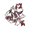
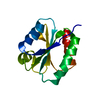
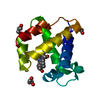
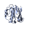
Structural insightsinto potent, specific anti-HIV property of actinohivin; Crystal structure of actinohivin in complex with alpha(1-2) mannobiose moiety of high-mannose type glycan of gp120
Components
Actinohivin
Keywords
ANTIVIRAL PROTEIN / anti-HIV lectin / molecular recognition / high-mannose type glycan
Resolution: 1.6→39.75 Å / Cor.coef. Fo:Fc: 0.972 / Cor.coef. Fo:Fc free: 0.928 / SU B: 1.914 / SU ML: 0.073 / Cross valid method: THROUGHOUT / ESU R: 0.126 / ESU R Free: 0.12 / Stereochemistry target values: MAXIMUM LIKELIHOOD Details: THE CRYSTAL HABIT SHOWS THE SPACE GROUP TO BE P213 APPARENTLY AT THE HIGHEST CONFIDENCE. IN THE CRYSTAL STRUCTURE, HOWEVER, AUTHORS HAVE FOUND THAT THREE ACTINOHIVIN MOLECULES (IN COMPLEX ...Details: THE CRYSTAL HABIT SHOWS THE SPACE GROUP TO BE P213 APPARENTLY AT THE HIGHEST CONFIDENCE. IN THE CRYSTAL STRUCTURE, HOWEVER, AUTHORS HAVE FOUND THAT THREE ACTINOHIVIN MOLECULES (IN COMPLEX WITH ALPHA(1-2)MANNOBIOSE) ARE DISORDERED AROUND THE CRYSTALLOGRAPHIC 3-FOLD AXIS. THIS IS ASCRIBED TO THE MOLECULAR PSEUDO 3-FOLD SYMMETRY OF THE INTRINSIC STRUCTURE. THEREFORE, IT IS CONCLUDED THAT ACTINOHIVIN MOLECULES ARE PACKED ACCORDING TO THE SPACE GROUP P212121, BUT THE PROTEINS CAN ROTATE RANDOMLY AT EVERY 120 DEGREE AROUND THE MOLECULAR SYMMETRY IN EACH UNIT CELL OR BETWEEN THE CELLS. THE DETAILED DATA AND DISCUSSIONS ARE DESCRIBED IN THE PUBLISHED PAPER.
Rfactor
Num. reflection
% reflection
Selection details
Rfree
0.20167
370
4.6 %
RANDOM
Rwork
0.14473
-
-
-
obs
0.14721
7647
99.16 %
-
Solvent computation
Ion probe radii: 0.8 Å / Shrinkage radii: 0.8 Å / VDW probe radii: 1.4 Å / Solvent model: MASK
Displacement parameters
Biso mean: 16.958 Å2
Refinement step
Cycle: LAST / Resolution: 1.6→39.75 Å
Protein
Nucleic acid
Ligand
Solvent
Total
Num. atoms
875
0
72
69
1016
Refine LS restraints
Refine-ID
Type
Dev ideal
Dev ideal target
Number
X-RAY DIFFRACTION
r_bond_refined_d
0.021
0.021
970
X-RAY DIFFRACTION
r_angle_refined_deg
2.197
2.003
1332
X-RAY DIFFRACTION
r_dihedral_angle_1_deg
6.728
5
111
X-RAY DIFFRACTION
r_dihedral_angle_2_deg
33.199
24.4
50
X-RAY DIFFRACTION
r_dihedral_angle_3_deg
15.246
15
121
X-RAY DIFFRACTION
r_dihedral_angle_4_deg
10.212
15
6
X-RAY DIFFRACTION
r_chiral_restr
0.153
0.2
152
X-RAY DIFFRACTION
r_gen_planes_refined
0.011
0.021
739
X-RAY DIFFRACTION
r_mcbond_it
1.176
1.5
547
X-RAY DIFFRACTION
r_mcangle_it
1.809
2
870
X-RAY DIFFRACTION
r_scbond_it
2.922
3
423
X-RAY DIFFRACTION
r_scangle_it
3.779
4.5
462
LS refinement shell
Resolution: 1.598→1.64 Å / Total num. of bins used: 20
Rfactor
Num. reflection
% reflection
Rfree
0.29
25
-
Rwork
0.197
554
-
obs
-
554
100 %
+
About Yorodumi
-
News
-
Feb 9, 2022. New format data for meta-information of EMDB entries
New format data for meta-information of EMDB entries
Version 3 of the EMDB header file is now the official format.
The previous official version 1.9 will be removed from the archive.
In the structure databanks used in Yorodumi, some data are registered as the other names, "COVID-19 virus" and "2019-nCoV". Here are the details of the virus and the list of structure data.
Jan 31, 2019. EMDB accession codes are about to change! (news from PDBe EMDB page)
EMDB accession codes are about to change! (news from PDBe EMDB page)
The allocation of 4 digits for EMDB accession codes will soon come to an end. Whilst these codes will remain in use, new EMDB accession codes will include an additional digit and will expand incrementally as the available range of codes is exhausted. The current 4-digit format prefixed with “EMD-” (i.e. EMD-XXXX) will advance to a 5-digit format (i.e. EMD-XXXXX), and so on. It is currently estimated that the 4-digit codes will be depleted around Spring 2019, at which point the 5-digit format will come into force.
The EM Navigator/Yorodumi systems omit the EMD- prefix.
Related info.:Q: What is EMD? / ID/Accession-code notation in Yorodumi/EM Navigator
Yorodumi is a browser for structure data from EMDB, PDB, SASBDB, etc.
This page is also the successor to EM Navigator detail page, and also detail information page/front-end page for Omokage search.
The word "yorodu" (or yorozu) is an old Japanese word meaning "ten thousand". "mi" (miru) is to see.
Related info.:EMDB / PDB / SASBDB / Comparison of 3 databanks / Yorodumi Search / Aug 31, 2016. New EM Navigator & Yorodumi / Yorodumi Papers / Jmol/JSmol / Function and homology information / Changes in new EM Navigator and Yorodumi
 Movie
Movie Controller

 Yorodumi
 Open data
 Basic information
 Components
 Keywords
 ANTIVIRAL PROTEIN / anti-HIV lectin /
  Function and homology information
 Actinomycete (bacteria)
 Authors
 Citation
 Structure visualization
 Downloads & links
 Other downloads
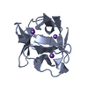


 PDBj
PDBj


 Assembly
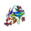


 Mass: 39.098 Da / Num. of mol.: 3 / Source method: obtained synthetically / Formula: K
 Mass: 18.015 Da / Num. of mol.: 69 / Source method: isolated from a natural source / Formula: H2O
 Sample preparation
 / Beamline: AR-NW12A / Wavelength: 1 Å
 Processing