 Movie
Movie Controller


+ Open data

- Basic information
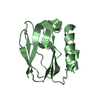
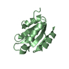
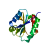
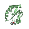
| Entry | Database: PDB / ID: 6ifp | ||||||
|---|---|---|---|---|---|---|---|
| Title | X-ray Crystal Structure of Pseudoazurin Met16Ile Variant | ||||||
 Components Components | Pseudoazurin | ||||||
 Keywords Keywords |  ELECTRON TRANSPORT ELECTRON TRANSPORT | ||||||
| Function / homology |  Function and homology information Function and homology informationelectron transporter, transferring electrons from cytochrome b6/f complex of photosystem II activity / periplasmic space / copper ion bindingSimilarity search - Function | ||||||
| Biological species |  Achromobacter cycloclastes (bacteria) Achromobacter cycloclastes (bacteria) | ||||||
| Method | X-RAY DIFFRACTION / SYNCHROTRON / MOLECULAR REPLACEMENT / molecular replacement / Resolution: 1 Å | ||||||
 Authors Authors | Sakai, C. / Yamaguchi, T. / Kohzuma, T. | ||||||
| Funding support |  Japan, 1items Japan, 1items
| ||||||
 Citation Citation | Journal: To Be Published Title: X-ray Crystal Structure of Pseudoazurin Met16Leu Variant Authors: Sakai, C. / Yamaguchi, T. / Kohzuma, T. | ||||||
| History |
|
- Structure visualization
| Structure viewer | Molecule: MolmilJmol/JSmol |
|---|
- Downloads & links
-Download
| PDBx/mmCIF format | 6ifp.cif.gz | 175.4 KB | Display | PDBx/mmCIF format |
|---|---|---|---|---|
| PDB format | pdb6ifp.ent.gz | 139.1 KB | Display | PDB format |
| PDBx/mmJSON format | 6ifp.json.gz | Tree view | PDBx/mmJSON format | |
| Others |  Other downloads Other downloads |
-Validation report
| Arichive directory | https://data.pdbj.org/pub/pdb/validation_reports/if/6ifpftp://data.pdbj.org/pub/pdb/validation_reports/if/6ifp | HTTPS FTP |
|---|
-Related structure data
| Related structure data |  6aknC  1bqkS S: Starting model for refinement C: citing same article ( |
|---|---|
| Similar structure data |


-Links
 PDBj
PDBj- Assembly
| Deposited unit | 
| ||||||||
|---|---|---|---|---|---|---|---|---|---|
| 1 | 
| ||||||||
| 2 | 
| ||||||||
| Unit cell |
|
-Components
| #1: Protein | Mass: 13014.902 Da / Num. of mol.: 2 / Fragment: UNP residues 29-152 / Mutation: M16I Source method: isolated from a genetically manipulated source Source: (gene. exp.) Achromobacter cycloclastes (bacteria) / Gene: bcp / Plasmid: pTrc99A / Production host: Escherichia coli (E. coli) / References: UniProt: P19567#2: Chemical | Copper  Mass: 63.546 Da / Num. of mol.: 2 / Source method: obtained synthetically / Formula: Cu Mass: 63.546 Da / Num. of mol.: 2 / Source method: obtained synthetically / Formula: Cu#3: Water | ChemComp-HOH / | Water Mass: 18.015 Da / Num. of mol.: 546 / Source method: isolated from a natural source / Formula: H2O Mass: 18.015 Da / Num. of mol.: 546 / Source method: isolated from a natural source / Formula: H2O |
|---|
-Experimental details
-Experiment
| Experiment | Method: X-RAY DIFFRACTION / Number of used crystals: 1 |
|---|
- Sample preparation
| Crystal | Density Matthews: 1.87 Å3/Da / Density % sol: 34.33 % |
|---|---|
| Crystal grow | Temperature: 298 K / Method: vapor diffusion, hanging drop / pH: 7.5 / Details: 0.1 M Tris-HCl, 35 % PEG4000 |
-Data collection
| Diffraction | Mean temperature: 100 K / Ambient temp details: cryostream / Serial crystal experiment: N | |||||||||||||||||||||||||||||||||||||||||||||||||||||||||||||||||||||||||||||||||||||||||||||||||||||||||||||||||||||||||||||||||||||||||||||||||||||||||||||||||||||||||||||||||||||||||||||
|---|---|---|---|---|---|---|---|---|---|---|---|---|---|---|---|---|---|---|---|---|---|---|---|---|---|---|---|---|---|---|---|---|---|---|---|---|---|---|---|---|---|---|---|---|---|---|---|---|---|---|---|---|---|---|---|---|---|---|---|---|---|---|---|---|---|---|---|---|---|---|---|---|---|---|---|---|---|---|---|---|---|---|---|---|---|---|---|---|---|---|---|---|---|---|---|---|---|---|---|---|---|---|---|---|---|---|---|---|---|---|---|---|---|---|---|---|---|---|---|---|---|---|---|---|---|---|---|---|---|---|---|---|---|---|---|---|---|---|---|---|---|---|---|---|---|---|---|---|---|---|---|---|---|---|---|---|---|---|---|---|---|---|---|---|---|---|---|---|---|---|---|---|---|---|---|---|---|---|---|---|---|---|---|---|---|---|---|---|---|---|
| Diffraction source | Source: SYNCHROTRON / Site: Photon Factory / Beamline: AR-NW12A / Wavelength: 1 Å | |||||||||||||||||||||||||||||||||||||||||||||||||||||||||||||||||||||||||||||||||||||||||||||||||||||||||||||||||||||||||||||||||||||||||||||||||||||||||||||||||||||||||||||||||||||||||||||
| Detector | Type: ADSC QUANTUM 270 / Detector: CCD / Date: Dec 18, 2017 | |||||||||||||||||||||||||||||||||||||||||||||||||||||||||||||||||||||||||||||||||||||||||||||||||||||||||||||||||||||||||||||||||||||||||||||||||||||||||||||||||||||||||||||||||||||||||||||
| Radiation | Protocol: SINGLE WAVELENGTH / Monochromatic (M) / Laue (L): M / Scattering type: x-ray | |||||||||||||||||||||||||||||||||||||||||||||||||||||||||||||||||||||||||||||||||||||||||||||||||||||||||||||||||||||||||||||||||||||||||||||||||||||||||||||||||||||||||||||||||||||||||||||
| Radiation wavelength | Wavelength: 1 Å / Relative weight: 1 | |||||||||||||||||||||||||||||||||||||||||||||||||||||||||||||||||||||||||||||||||||||||||||||||||||||||||||||||||||||||||||||||||||||||||||||||||||||||||||||||||||||||||||||||||||||||||||||
| Reflection | Resolution: 1→50 Å / Num. obs: 103433 / % possible obs: 100 % / Redundancy: 7.1 % / Rmerge(I) obs: 0.092 / Rpim(I) all: 0.037 / Rrim(I) all: 0.099 / Χ2: 1.826 / Net I/σ(I): 8 / Num. measured all: 737381 | |||||||||||||||||||||||||||||||||||||||||||||||||||||||||||||||||||||||||||||||||||||||||||||||||||||||||||||||||||||||||||||||||||||||||||||||||||||||||||||||||||||||||||||||||||||||||||||
| Reflection shell | Diffraction-ID: 1
|
-Phasing
| Phasing | Method: molecular replacement | ||||||
|---|---|---|---|---|---|---|---|
| Phasing MR | R rigid body: 0.661
|
- Processing
| Software |
| ||||||||||||||||||||||||
|---|---|---|---|---|---|---|---|---|---|---|---|---|---|---|---|---|---|---|---|---|---|---|---|---|---|
| Refinement | Method to determine structure: MOLECULAR REPLACEMENT Starting model: 1BQK Resolution: 1→30 Å / Cross valid method: FREE R-VALUE
| ||||||||||||||||||||||||
| Displacement parameters | Biso max: 112.58 Å2 / Biso mean: 15.1194 Å2 / Biso min: 3.37 Å2 | ||||||||||||||||||||||||
| Refinement step | Cycle: LAST / Resolution: 1→30 Å
| ||||||||||||||||||||||||
| LS refinement shell | Resolution: 1→1.04 Å /
|
