 Movie
Movie Controller

[English] 日本語
 Yorodumi
- PDB-4d62: Structure of the carboxy-terminal domain of the turkey type 3 sia... -
+ Open data

- Basic information
| Entry | Database: PDB / ID: 4d62 | ||||||
|---|---|---|---|---|---|---|---|
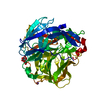
| Title | Structure of the carboxy-terminal domain of the turkey type 3 siadenovirus fibre, avirulent form complexed with 3-sialyllactose. | ||||||
 Components Components | FIBER KNOB DOMAIN | ||||||
 Keywords Keywords |  VIRAL PROTEIN VIRAL PROTEIN | ||||||
| Function / homology |  Function and homology information Function and homology informationsymbiont entry into host cell / host cell nucleus / virion attachment to host cell Similarity search - Function | ||||||
| Biological species |  AVIRULENT TURKEY HEMORRHAGIC ENTERITIS VIRUS AVIRULENT TURKEY HEMORRHAGIC ENTERITIS VIRUS | ||||||
| Method | X-RAY DIFFRACTION / SYNCHROTRON / OTHER / Resolution: 2.5 Å | ||||||
 Authors Authors | Singh, A.K. / van Raaij, M.J. | ||||||
 Citation Citation | Journal: PLoS ONE / Year: 2015 Title: Structure and Sialyllactose Binding of the Carboxy-Terminal Head Domain of the Fibre from a Siadenovirus, Turkey Adenovirus 3. Authors: Singh, A.K. / Berbis, M.A. / Ballmann, M.Z. / Kilcoyne, M. / Menendez, M. / Nguyen, T.H. / Joshi, L. / Canada, F.J. / Jimenez-Barbero, J. / Benko, M. / Harrach, B. / van Raaij, M.J. | ||||||
| History |
|
- Structure visualization
| Structure viewer | Molecule: MolmilJmol/JSmol |
|---|
- Downloads & links
-Download
| PDBx/mmCIF format | 4d62.cif.gz | 42.5 KB | Display | PDBx/mmCIF format |
|---|---|---|---|---|
| PDB format | pdb4d62.ent.gz | 29.3 KB | Display | PDB format |
| PDBx/mmJSON format | 4d62.json.gz | Tree view | PDBx/mmJSON format | |
| Others |  Other downloads Other downloads |
-Validation report
| Arichive directory | https://data.pdbj.org/pub/pdb/validation_reports/d6/4d62ftp://data.pdbj.org/pub/pdb/validation_reports/d6/4d62 | HTTPS FTP |
|---|
-Related structure data


-Links
 PDBj
PDBj

- Assembly
| Deposited unit | 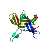
| ||||||||
|---|---|---|---|---|---|---|---|---|---|
| 1 | 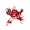
| ||||||||
| Unit cell |
|
-Components
| #1: Protein | Mass: 20611.973 Da / Num. of mol.: 1 / Fragment: HEAD DOMAIN, RESIDUES 15-165 Source method: isolated from a genetically manipulated source Source: (gene. exp.) AVIRULENT TURKEY HEMORRHAGIC ENTERITIS VIRUSDescription: ARKO LABORATORIES, JEWELL IA, USA / Variant: AVIRULENT FORM / Plasmid: PET28A / Production host:  ESCHERICHIA COLI (E. coli) / Strain (production host): BL21(DE3) / References: UniProt: Q2TLC1 ESCHERICHIA COLI (E. coli) / Strain (production host): BL21(DE3) / References: UniProt: Q2TLC1 |
|---|---|
| #2: Sugar | ChemComp-SIA / Sialic acid  Type: D-saccharide, alpha linking / Mass: 309.270 Da / Num. of mol.: 1 Type: D-saccharide, alpha linking / Mass: 309.270 Da / Num. of mol.: 1Source method: isolated from a genetically manipulated source Formula: C11H19NO9 |
| #3: Water | ChemComp-HOH / Water Mass: 18.015 Da / Num. of mol.: 18 / Source method: isolated from a natural source / Formula: H2O Mass: 18.015 Da / Num. of mol.: 18 / Source method: isolated from a natural source / Formula: H2O |
-Experimental details
-Experiment
| Experiment | Method: X-RAY DIFFRACTION / Number of used crystals: 1 |
|---|
- Sample preparation
| Crystal | Density Matthews: 1.94 Å3/Da / Density % sol: 37 % / Description: NONE |
|---|---|
| Crystal grow | pH: 5.5 Details: 25 MM MORPHOLINO-ETHANESULFONIC ACID, 0.5-1.0M DIAMMONIUM PHOSPHATE, 0.1M SODIUM CITRATE PH 5.5-5.8, 0.2-0.3 M SODIUM CHLORIDE |
-Data collection
| Diffraction | Mean temperature: 100 K |
|---|---|
| Diffraction source | Source: SYNCHROTRON / Site: ESRF  / Beamline: BM30A / Wavelength: 0.9797 / Beamline: BM30A / Wavelength: 0.9797 |
| Detector | Type: ADSC QUANTUM 315r / Detector: CCD / Date: Jul 11, 2013 Details: TWO CYLINDRICAL VERTICAL FOCUSING PARABOLIC MIRRORS |
| Radiation | Monochromator: SI (111), SI(311) DOUBLE CRYSTAL MONOCHROMATOR Protocol: SINGLE WAVELENGTH / Monochromatic (M) / Laue (L): M / Scattering type: x-ray |
| Radiation wavelength | Wavelength: 0.9797 Å / Relative weight: 1 |
| Reflection | Resolution: 2.5→25 Å / Num. obs: 5355 / % possible obs: 100 % / Redundancy: 7.7 % / Biso Wilson estimate: 44.6 Å2 / Rmerge(I) obs: 0.1 / Net I/σ(I): 14.6 |
| Reflection shell | Resolution: 2.5→2.64 Å / Redundancy: 7.9 % / Rmerge(I) obs: 0.96 / Mean I/σ(I) obs: 2.6 / % possible all: 100 |
- Processing
| Software |
| ||||||||||||||||||||||||||||||||||||||||||||||||||||||||||||||||||||||||||||||||||||||||||||||||||||||||||||||||||||||||||||||||||||||||||||||||||||||||||||||||||||||||||||||||||||||
|---|---|---|---|---|---|---|---|---|---|---|---|---|---|---|---|---|---|---|---|---|---|---|---|---|---|---|---|---|---|---|---|---|---|---|---|---|---|---|---|---|---|---|---|---|---|---|---|---|---|---|---|---|---|---|---|---|---|---|---|---|---|---|---|---|---|---|---|---|---|---|---|---|---|---|---|---|---|---|---|---|---|---|---|---|---|---|---|---|---|---|---|---|---|---|---|---|---|---|---|---|---|---|---|---|---|---|---|---|---|---|---|---|---|---|---|---|---|---|---|---|---|---|---|---|---|---|---|---|---|---|---|---|---|---|---|---|---|---|---|---|---|---|---|---|---|---|---|---|---|---|---|---|---|---|---|---|---|---|---|---|---|---|---|---|---|---|---|---|---|---|---|---|---|---|---|---|---|---|---|---|---|---|---|
| Refinement | Method to determine structure: OTHER Starting model: NONE Resolution: 2.5→25 Å / Cor.coef. Fo:Fc: 0.956 / Cor.coef. Fo:Fc free: 0.911 / SU B: 12.19 / SU ML: 0.256 / Cross valid method: THROUGHOUT / ESU R: 0.574 / ESU R Free: 0.297 / Stereochemistry target values: MAXIMUM LIKELIHOOD Details: HYDROGENS HAVE BEEN ADDED IN THE RIDING POSITIONS. U VALUES REFINED INDIVIDUALLY
| ||||||||||||||||||||||||||||||||||||||||||||||||||||||||||||||||||||||||||||||||||||||||||||||||||||||||||||||||||||||||||||||||||||||||||||||||||||||||||||||||||||||||||||||||||||||
| Solvent computation | Ion probe radii: 0.8 Å / Shrinkage radii: 0.8 Å / VDW probe radii: 1.2 Å / Solvent model: MASK | ||||||||||||||||||||||||||||||||||||||||||||||||||||||||||||||||||||||||||||||||||||||||||||||||||||||||||||||||||||||||||||||||||||||||||||||||||||||||||||||||||||||||||||||||||||||
| Displacement parameters | Biso mean: 52.906 Å2
| ||||||||||||||||||||||||||||||||||||||||||||||||||||||||||||||||||||||||||||||||||||||||||||||||||||||||||||||||||||||||||||||||||||||||||||||||||||||||||||||||||||||||||||||||||||||
| Refinement step | Cycle: LAST / Resolution: 2.5→25 Å
| ||||||||||||||||||||||||||||||||||||||||||||||||||||||||||||||||||||||||||||||||||||||||||||||||||||||||||||||||||||||||||||||||||||||||||||||||||||||||||||||||||||||||||||||||||||||
| Refine LS restraints |
|