 Movie
Movie Controller

[English] 日本語
 Yorodumi
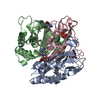
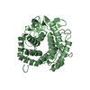
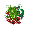
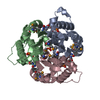
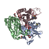
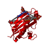
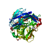
- PDB-3ern: Crystal structure of 2C-methyl-D-erythritol 2,4-clycodiphosphate ... -
+ Open data

- Basic information
| Entry | Database: PDB / ID: 3ern | ||||||
|---|---|---|---|---|---|---|---|
| Title | Crystal structure of 2C-methyl-D-erythritol 2,4-clycodiphosphate synthase complexed with AraCMP | ||||||
 Components Components | 2-C-methyl-D-erythritol 2,4-cyclodiphosphate synthase | ||||||
 Keywords Keywords | LYASE / MECDP-synthase / Isoprene biosynthesis / Magnesium / Manganese / Metal-binding | ||||||
| Function / homology |  Function and homology information2-C-methyl-D-erythritol 2,4-cyclodiphosphate synthase / 2-C-methyl-D-erythritol 2,4-cyclodiphosphate synthase activity / ubiquinone biosynthetic process / isopentenyl diphosphate biosynthetic process, methylerythritol 4-phosphate pathway / terpenoid biosynthetic process / manganese ion binding / zinc ion binding / identical protein binding / metal ion binding Function and homology information2-C-methyl-D-erythritol 2,4-cyclodiphosphate synthase / 2-C-methyl-D-erythritol 2,4-cyclodiphosphate synthase activity / ubiquinone biosynthetic process / isopentenyl diphosphate biosynthetic process, methylerythritol 4-phosphate pathway / terpenoid biosynthetic process / manganese ion binding / zinc ion binding / identical protein binding / metal ion bindingSimilarity search - Function | ||||||
| Biological species |  Escherichia coli K-12 (bacteria) Escherichia coli K-12 (bacteria) | ||||||
| Method | X-RAY DIFFRACTION / SYNCHROTRON / MOLECULAR REPLACEMENT / Resolution: 2.1 Å | ||||||
 Authors Authors | Hunter, W.N. / Ramsden, N.L. / Kemp, L.A. | ||||||
 Citation Citation | Journal: J.Med.Chem. / Year: 2009 Title: A structure-based approach to ligand discovery for 2C-methyl-D-erythritol-2,4-cyclodiphosphate synthase: a target for antimicrobial therapy Authors: Ramsden, N.L. / Buetow, L. / Dawson, A. / Kemp, L.A. / Ulaganathan, V. / Brenk, R. / Klebe, G. / Hunter, W.N. | ||||||
| History |
|
- Structure visualization
| Structure viewer | Molecule: MolmilJmol/JSmol |
|---|
- Downloads & links
-Download
| PDBx/mmCIF format | 3ern.cif.gz | 192.4 KB | Display | PDBx/mmCIF format |
|---|---|---|---|---|
| PDB format | pdb3ern.ent.gz | 154.1 KB | Display | PDB format |
| PDBx/mmJSON format | 3ern.json.gz | Tree view | PDBx/mmJSON format | |
| Others |  Other downloads Other downloads |
-Validation report
| Arichive directory | https://data.pdbj.org/pub/pdb/validation_reports/er/3ernftp://data.pdbj.org/pub/pdb/validation_reports/er/3ern | HTTPS FTP |
|---|
-Related structure data
| Related structure data |  3elcC  3eorC  3esjC  3fbaC  1gx1S S: Starting model for refinement C: citing same article ( |
|---|---|
| Similar structure data |
-Links
 PDBj
PDBj- Assembly
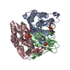
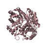
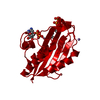
| Deposited unit | 
| ||||||||
|---|---|---|---|---|---|---|---|---|---|
| 1 | 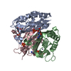
| ||||||||
| 2 | 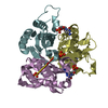
| ||||||||
| Unit cell |
|
-Components
-Protein , 1 types, 6 molecules ABCDEF
| #1: Protein | / MECPS / MECDP-synthase Mass: 17576.275 Da / Num. of mol.: 6 Source method: isolated from a genetically manipulated source Source: (gene. exp.) Escherichia coli K-12 (bacteria) / Strain: K12 / Gene: ispF / Plasmid: pET15b / Production host: Escherichia coli BL21(DE3) (bacteria) / Strain (production host): BL21(DE3)References: UniProt: P62617, 2-C-methyl-D-erythritol 2,4-cyclodiphosphate synthase |
|---|
-Non-polymers , 5 types, 297 molecules 


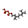

| #2: Chemical | ChemComp-ZN /  Mass: 65.409 Da / Num. of mol.: 4 / Source method: obtained synthetically / Formula: Zn Mass: 65.409 Da / Num. of mol.: 4 / Source method: obtained synthetically / Formula: Zn#3: Chemical | ChemComp-CAR /  Type: RNA linking / Mass: 323.197 Da / Num. of mol.: 5 / Source method: obtained synthetically / Formula: C9H14N3O8P Type: RNA linking / Mass: 323.197 Da / Num. of mol.: 5 / Source method: obtained synthetically / Formula: C9H14N3O8P#4: Chemical | Sulfate Mass: 96.063 Da / Num. of mol.: 2 / Source method: obtained synthetically / Formula: SO4 Mass: 96.063 Da / Num. of mol.: 2 / Source method: obtained synthetically / Formula: SO4#5: Chemical | Geranyl pyrophosphate Mass: 314.209 Da / Num. of mol.: 2 / Source method: obtained synthetically / Formula: C10H20O7P2 Mass: 314.209 Da / Num. of mol.: 2 / Source method: obtained synthetically / Formula: C10H20O7P2#6: Water | ChemComp-HOH / | WaterMass: 18.015 Da / Num. of mol.: 284 / Source method: isolated from a natural source / Formula: H2O |
|---|
-Experimental details
-Experiment
| Experiment | Method: X-RAY DIFFRACTION / Number of used crystals: 1 |
|---|
- Sample preparation
| Crystal | Density Matthews: 2.69 Å3/Da / Density % sol: 54.35 % |
|---|---|
| Crystal grow | Temperature: 298 K / Method: vapor diffusion, hanging drop / pH: 4.4 Details: 0.1M ammonium sulfate, sodium acetate, PEG 2000 MME, pH 4.4, vapor diffusion, hanging drop, temperature 298K |
-Data collection
| Diffraction | Mean temperature: 100 K |
|---|---|
| Diffraction source | Source: SYNCHROTRON / Site: ESRF  / Beamline: ID29 / Wavelength: 0.9756 Å / Beamline: ID29 / Wavelength: 0.9756 Å |
| Detector | Type: ADSC QUANTUM 210 / Detector: CCD / Date: Nov 1, 2003 / Details: mirrors |
| Radiation | Monochromator: SILICON / Protocol: SINGLE WAVELENGTH / Monochromatic (M) / Laue (L): M / Scattering type: x-ray |
| Radiation wavelength | Wavelength: 0.9756 Å / Relative weight: 1 |
| Reflection | Resolution: 2.1→20 Å / Num. all: 71490 / Num. obs: 65677 / % possible obs: 91.86 % / Observed criterion σ(F): 0 / Observed criterion σ(I): 1.6 / Redundancy: 4.6 % / Rsym value: 0.089 |
| Reflection shell | Resolution: 2.1→2.154 Å / % possible all: 92.95 |
- Processing
| Software |
| ||||||||||||||||||||||||||||||||||||||||||||||||||||||||||||||||||||||||||||||||||||||||||
|---|---|---|---|---|---|---|---|---|---|---|---|---|---|---|---|---|---|---|---|---|---|---|---|---|---|---|---|---|---|---|---|---|---|---|---|---|---|---|---|---|---|---|---|---|---|---|---|---|---|---|---|---|---|---|---|---|---|---|---|---|---|---|---|---|---|---|---|---|---|---|---|---|---|---|---|---|---|---|---|---|---|---|---|---|---|---|---|---|---|---|---|
| Refinement | Method to determine structure: MOLECULAR REPLACEMENT Starting model: 1GX1 Resolution: 2.1→20 Å / Cor.coef. Fo:Fc: 0.945 / Cor.coef. Fo:Fc free: 0.933 / WRfactor Rfree: 0.223 / WRfactor Rwork: 0.207 / Occupancy max: 1 / Occupancy min: 0 / FOM work R set: 0.806 / SU B: 5.691 / SU ML: 0.148 / SU R Cruickshank DPI: 0.222 / SU Rfree: 0.175 / Cross valid method: THROUGHOUT / σ(F): 0 / ESU R: 0.222 / ESU R Free: 0.175 / Stereochemistry target values: MAXIMUM LIKELIHOOD
| ||||||||||||||||||||||||||||||||||||||||||||||||||||||||||||||||||||||||||||||||||||||||||
| Solvent computation | Ion probe radii: 0.8 Å / Shrinkage radii: 0.8 Å / VDW probe radii: 1.2 Å / Solvent model: BABINET MODEL WITH MASK | ||||||||||||||||||||||||||||||||||||||||||||||||||||||||||||||||||||||||||||||||||||||||||
| Displacement parameters | Biso max: 96.14 Å2 / Biso mean: 34.02 Å2 / Biso min: 15.45 Å2
| ||||||||||||||||||||||||||||||||||||||||||||||||||||||||||||||||||||||||||||||||||||||||||
| Refinement step | Cycle: LAST / Resolution: 2.1→20 Å
| ||||||||||||||||||||||||||||||||||||||||||||||||||||||||||||||||||||||||||||||||||||||||||
| Refine LS restraints |
| ||||||||||||||||||||||||||||||||||||||||||||||||||||||||||||||||||||||||||||||||||||||||||
| LS refinement shell | Resolution: 2.1→2.154 Å / Total num. of bins used: 20
|
