 Movie
Movie Controller

[English] 日本語
 Yorodumi
- PDB-1h21: A novel iron centre in the split-Soret cytochrome c from Desulfov... -
+ Open data

- Basic information
| Entry | Database: PDB / ID: 1h21 | |||||||||
|---|---|---|---|---|---|---|---|---|---|---|
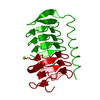
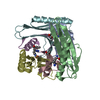
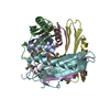
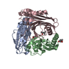
| Title | A novel iron centre in the split-Soret cytochrome c from Desulfovibrio desulfuricans ATCC 27774 | |||||||||
 Components Components | SPLIT-SORET CYTOCHROME C | |||||||||
 Keywords Keywords |  CYTOCHROME / DIMERIC DI-HEME CYTOCHROME / STACKED HEME ARRANGEMENT / NOVEL FOLD / NOVEL IRON-SULFUR CENTRE / ELECTRON TRANSPORT / SULFATE RESPIRATION CYTOCHROME / DIMERIC DI-HEME CYTOCHROME / STACKED HEME ARRANGEMENT / NOVEL FOLD / NOVEL IRON-SULFUR CENTRE / ELECTRON TRANSPORT / SULFATE RESPIRATION | |||||||||
| Function / homology |  Function and homology informationanaerobic respiration / 2 iron, 2 sulfur cluster binding / metal ion binding Function and homology informationanaerobic respiration / 2 iron, 2 sulfur cluster binding / metal ion bindingSimilarity search - Function | |||||||||
| Biological species |  DESULFOVIBRIO DESULFURICANS (bacteria) DESULFOVIBRIO DESULFURICANS (bacteria) | |||||||||
| Method | X-RAY DIFFRACTION / SYNCHROTRON / MAD / Resolution: 2.5 Å | |||||||||
 Authors Authors | Abreu, I.A. / Lourenco, A.I. / Xavier, A.V. / Legall, J. / Coelho, A.V. / Matias, P.M. / Pinto, D.M. / Carrondo, M.A. / Teixeira, M. / Saraiva, L.M. | |||||||||
 Citation Citation | Journal: J.Biol.Inorg.Chem. / Year: 2003 Title: A Novel Iron Centre in the Split-Soret Cytochrome C from Desulfovibrio Desulfuricans Atcc 27774 Authors: Abreu, I.A. / Lourenco, A.I. / Xavier, A.V. / Legall, J. / Coelho, A.V. / Matias, P.M. / Pinto, D.M. / Armenia Carrondo, M. / Teixeira, M. / Saraiva, L.M. #1: Journal: J.Biol.Inorg.Chem. / Year: 1997Title: A Preliminary Analysis of the Three Dimensional Structure of Dimeric Di-Haem Split-Soret Cytochrome C from Desulfovibrio Desulfuricans Atcc 27774 at 2.5 A Resolution Using the MAD Phasing ...Title: A Preliminary Analysis of the Three Dimensional Structure of Dimeric Di-Haem Split-Soret Cytochrome C from Desulfovibrio Desulfuricans Atcc 27774 at 2.5 A Resolution Using the MAD Phasing Method: A Novel Cytochrome Fold with a Stacked Haem Arrangement Authors: Matias, P.M. / Morais, J. / Coelho, A.V. / Meijers, R. / Gonzalez, A. / Thompson, A.W. / Sieker, L. / Legall, J. / Carrondo, M.A. | |||||||||
| History |
|
- Structure visualization
| Structure viewer | Molecule: MolmilJmol/JSmol |
|---|
- Downloads & links
-Download
| PDBx/mmCIF format | 1h21.cif.gz | 197.9 KB | Display | PDBx/mmCIF format |
|---|---|---|---|---|
| PDB format | pdb1h21.ent.gz | 169.3 KB | Display | PDB format |
| PDBx/mmJSON format | 1h21.json.gz | Tree view | PDBx/mmJSON format | |
| Others |  Other downloads Other downloads |
-Validation report
| Arichive directory | https://data.pdbj.org/pub/pdb/validation_reports/h2/1h21ftp://data.pdbj.org/pub/pdb/validation_reports/h2/1h21 | HTTPS FTP |
|---|
-Related structure data
| Similar structure data |
|---|


-Links
 PDBj
PDBj


- Assembly
| Deposited unit | 
| ||||||||||||
|---|---|---|---|---|---|---|---|---|---|---|---|---|---|
| 1 | 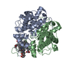
| ||||||||||||
| 2 | 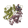
| ||||||||||||
| Unit cell |
| ||||||||||||
| Noncrystallographic symmetry (NCS) | NCS oper:
|
-Components
| #1: Protein | Mass: 26939.469 Da / Num. of mol.: 4 / Source method: isolated from a natural source / Source: (natural) DESULFOVIBRIO DESULFURICANS (bacteria) / References: UniProt: P81040#2: Chemical | ChemComp-HEC / Heme C  Mass: 618.503 Da / Num. of mol.: 8 / Source method: obtained synthetically / Formula: C34H34FeN4O4 Mass: 618.503 Da / Num. of mol.: 8 / Source method: obtained synthetically / Formula: C34H34FeN4O4#3: Water | ChemComp-HOH / | Water Mass: 18.015 Da / Num. of mol.: 188 / Source method: isolated from a natural source / Formula: H2O Mass: 18.015 Da / Num. of mol.: 188 / Source method: isolated from a natural source / Formula: H2OSequence details | GENBANK ENTRY AF465622 IS ON HOLD PENDING MANUSCRIPT ACCEPTANCE FOR PUBLICATION. SEQADV RECORDS ...GENBANK ENTRY AF465622 IS ON HOLD PENDING MANUSCRIPT | |
|---|
-Experimental details
-Experiment
| Experiment | Method: X-RAY DIFFRACTION / Number of used crystals: 1 |
|---|
- Sample preparation
| Crystal | Density Matthews: 2.59 Å3/Da / Density % sol: 53 % | ||||||||||||||||||||||||||||||||||||||||||
|---|---|---|---|---|---|---|---|---|---|---|---|---|---|---|---|---|---|---|---|---|---|---|---|---|---|---|---|---|---|---|---|---|---|---|---|---|---|---|---|---|---|---|---|
| Crystal grow | pH: 5 Details: PROTEIN WAS CRYSTALLIZED FROM A SOLUTION CONTAINING 0.1-0.2 % (W/V) AGAROSE IN 12-15 % PEG 8K, 0.1M SODIUM ACETATE BUFFER PH 5.0 | ||||||||||||||||||||||||||||||||||||||||||
| Crystal grow | *PLUS Method: vapor diffusion, sitting drop / Details: Matias, P.M., (1997) J.Biol.Inorg.Chem., 2, 507. | ||||||||||||||||||||||||||||||||||||||||||
| Components of the solutions | *PLUS
|
-Data collection
| Diffraction | Mean temperature: 100 K |
|---|---|
| Diffraction source | Source: SYNCHROTRON / Site: ESRF  / Beamline: BM14 / Wavelength: 1.7401 / Beamline: BM14 / Wavelength: 1.7401 |
| Detector | Type: MARRESEARCH / Detector: IMAGE PLATE / Date: Feb 15, 1996 / Details: MIRRORS |
| Radiation | Monochromator: SI (111) / Protocol: MAD / Monochromatic (M) / Laue (L): M / Scattering type: x-ray |
| Radiation wavelength | Wavelength: 1.7401 Å / Relative weight: 1 |
| Reflection | Resolution: 2.5→20 Å / Num. obs: 34228 / % possible obs: 93 % / Redundancy: 6.2 % / Rmerge(I) obs: 0.038 / Net I/σ(I): 9.7 |
| Reflection shell | Resolution: 2.5→2.56 Å / Rmerge(I) obs: 0.074 / Mean I/σ(I) obs: 9.7 / % possible all: 86 |
- Processing
| Software |
| ||||||||||||||||||||||||||||||||||||||||||||||||||||||||||||
|---|---|---|---|---|---|---|---|---|---|---|---|---|---|---|---|---|---|---|---|---|---|---|---|---|---|---|---|---|---|---|---|---|---|---|---|---|---|---|---|---|---|---|---|---|---|---|---|---|---|---|---|---|---|---|---|---|---|---|---|---|---|
| Refinement | Method to determine structure: MAD / Resolution: 2.5→20 Å / Data cutoff high absF: 100000 / Data cutoff low absF: 0.1 / Isotropic thermal model: RESTRAINED / Cross valid method: THROUGHOUT / σ(F): 2 / Details: FIRST 7 RESIDUES NOT VISIBLE IN ELECTRON DENSITY.
| ||||||||||||||||||||||||||||||||||||||||||||||||||||||||||||
| Displacement parameters | Biso mean: 35.04 Å2 | ||||||||||||||||||||||||||||||||||||||||||||||||||||||||||||
| Refine analyze | Luzzati d res low obs: 10 Å / Luzzati sigma a obs: 0.35 Å | ||||||||||||||||||||||||||||||||||||||||||||||||||||||||||||
| Refinement step | Cycle: LAST / Resolution: 2.5→20 Å
| ||||||||||||||||||||||||||||||||||||||||||||||||||||||||||||
| Refine LS restraints |
| ||||||||||||||||||||||||||||||||||||||||||||||||||||||||||||
| Xplor file |
| ||||||||||||||||||||||||||||||||||||||||||||||||||||||||||||
| Refine LS restraints | *PLUS
|
