 Movie
Movie Controller

[English] 日本語
 Yorodumi
- PDB-1mt1: The Crystal Structure of Pyruvoyl-dependent Arginine Decarboxylas... -
+ Open data

- Basic information
| Entry | Database: PDB / ID: 1mt1 | |||||||||
|---|---|---|---|---|---|---|---|---|---|---|
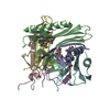
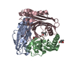
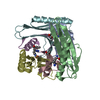
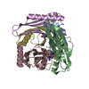
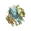
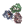
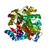
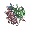
| Title | The Crystal Structure of Pyruvoyl-dependent Arginine Decarboxylase from Methanococcus jannaschii | |||||||||
 Components Components |
| |||||||||
 Keywords Keywords |  LYASE / pyruvoyl group / pyruvate / agmatine / arginine LYASE / pyruvoyl group / pyruvate / agmatine / arginine | |||||||||
| Function / homology |  Function and homology informationarginine decarboxylase / arginine decarboxylase activity / arginine catabolic process Function and homology informationarginine decarboxylase / arginine decarboxylase activity / arginine catabolic processSimilarity search - Function | |||||||||
| Biological species |   Methanocaldococcus jannaschii (archaea) Methanocaldococcus jannaschii (archaea) | |||||||||
| Method | X-RAY DIFFRACTION / SYNCHROTRON / SAD / Resolution: 2.2 Å | |||||||||
 Authors Authors | Tolbert, W.D. / Graham, D.E. / White, R.H. / Ealick, S.E. | |||||||||
 Citation Citation | Journal: Structure / Year: 2003 Title: Pyruvoyl-Dependent Arginine Decarboxylase from Methanococcus jannaschii: Crystal Structures of the Self-Cleaved and S53A Proenzyme Forms Authors: Tolbert, W.D. / Graham, D.E. / White, R.H. / Ealick, S.E. #1: Journal: J.Biol.Chem. / Year: 2002Title: Methanococcus jannaschii uses a pyruvoyl-dependent arginine decarboxylase in polyamine biosynthesis. Authors: Graham, D.E. / Xu, H. / White, R.H. | |||||||||
| History |
| |||||||||
| Remark 999 | SEQUENCE Each monomer undergoes an internal self cleavage reaction which generates a pyruvoyl ...SEQUENCE Each monomer undergoes an internal self cleavage reaction which generates a pyruvoyl cofactor. RESIDUE 53 IS CONVERTED TO A PYRUVOYL GROUP. |
- Structure visualization
| Structure viewer | Molecule: MolmilJmol/JSmol |
|---|
- Downloads & links
-Download
| PDBx/mmCIF format | 1mt1.cif.gz | 204.7 KB | Display | PDBx/mmCIF format |
|---|---|---|---|---|
| PDB format | pdb1mt1.ent.gz | 165.9 KB | Display | PDB format |
| PDBx/mmJSON format | 1mt1.json.gz | Tree view | PDBx/mmJSON format | |
| Others |  Other downloads Other downloads |
-Validation report
| Arichive directory | https://data.pdbj.org/pub/pdb/validation_reports/mt/1mt1ftp://data.pdbj.org/pub/pdb/validation_reports/mt/1mt1 | HTTPS FTP |
|---|
-Related structure data


-Links
 PDBj
PDBj- Assembly
| Deposited unit | 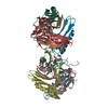
| ||||||||
|---|---|---|---|---|---|---|---|---|---|
| 1 | 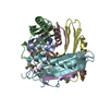
| ||||||||
| 2 | 
| ||||||||
| Unit cell |
| ||||||||
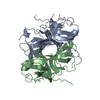
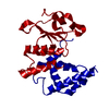
| Details | Two (alpha beta)3 trimers are located in the asymmetric unit. Each monomer undergoes an internal self cleavage reaction which generates a pyruvoyl cofactor. |
-Components
| #1: Protein | Mass: 5475.011 Da / Num. of mol.: 6 Source method: isolated from a genetically manipulated source Source: (gene. exp.) Methanocaldococcus jannaschii (archaea)Gene: MJ0316 / Plasmid: pET19b (Novagen) / Production host:  Escherichia coli (E. coli) Escherichia coli (E. coli)Strain (production host): BL21-CodonPlus(DE3)-RIL (Stratagene) References: UniProt: Q57764, arginine decarboxylase#2: Protein | Mass: 12531.892 Da / Num. of mol.: 6 Source method: isolated from a genetically manipulated source Source: (gene. exp.) Methanocaldococcus jannaschii (archaea)Gene: MJ0316 / Plasmid: pET19b (Novagen) / Production host: Escherichia coli (E. coli)Strain (production host): BL21-CodonPlus(DE3)-RIL (Stratagene) References: UniProt: Q57764, arginine decarboxylase#3: Chemical | ChemComp-AG2 / Agmatine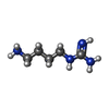  Mass: 130.191 Da / Num. of mol.: 5 / Source method: obtained synthetically / Formula: C5H14N4 Mass: 130.191 Da / Num. of mol.: 5 / Source method: obtained synthetically / Formula: C5H14N4#4: Water | ChemComp-HOH / | Water Mass: 18.015 Da / Num. of mol.: 454 / Source method: isolated from a natural source / Formula: H2O Mass: 18.015 Da / Num. of mol.: 454 / Source method: isolated from a natural source / Formula: H2O |
|---|
-Experimental details
-Experiment
| Experiment | Method: X-RAY DIFFRACTION / Number of used crystals: 1 |
|---|
- Sample preparation
| Crystal | Density Matthews: 2.12 Å3/Da / Density % sol: 42.09 % | ||||||||||||||||||||||||||||||||||||||||||||||||||||||
|---|---|---|---|---|---|---|---|---|---|---|---|---|---|---|---|---|---|---|---|---|---|---|---|---|---|---|---|---|---|---|---|---|---|---|---|---|---|---|---|---|---|---|---|---|---|---|---|---|---|---|---|---|---|---|---|
| Crystal grow | Temperature: 298 K / Method: vapor diffusion, hanging drop / pH: 7 Details: PEG 2000, 2-methyl-2,4-pentanediol, glycerol, n-[2-hydroxyethyl]piperazine-N'-[2-ethanesulfonic acid], beta octyl glucoside, putrescine, ethylenediaminetetraacetic acid dithiothreitol, pH 7. ...Details: PEG 2000, 2-methyl-2,4-pentanediol, glycerol, n-[2-hydroxyethyl]piperazine-N'-[2-ethanesulfonic acid], beta octyl glucoside, putrescine, ethylenediaminetetraacetic acid dithiothreitol, pH 7.0, VAPOR DIFFUSION, HANGING DROP, temperature 298K | ||||||||||||||||||||||||||||||||||||||||||||||||||||||
| Crystal grow | *PLUS | ||||||||||||||||||||||||||||||||||||||||||||||||||||||
| Components of the solutions | *PLUS
|
-Data collection
| Diffraction | Mean temperature: 100 K |
|---|---|
| Diffraction source | Source: SYNCHROTRON / Site: APS  / Beamline: 8-BM / Wavelength: 0.9793 Å / Beamline: 8-BM / Wavelength: 0.9793 Å |
| Detector | Type: ADSC QUANTUM 315 / Detector: CCD / Date: Apr 18, 2002 |
| Radiation | Monochromator: Si 111 / Protocol: SINGLE WAVELENGTH / Monochromatic (M) / Laue (L): M / Scattering type: x-ray |
| Radiation wavelength | Wavelength: 0.9793 Å / Relative weight: 1 |
| Reflection | Resolution: 2.11→63.5 Å / Num. all: 57557 / Num. obs: 57557 / % possible obs: 99.9 % / Observed criterion σ(F): 0 / Observed criterion σ(I): 0 / Redundancy: 6.9 % / Biso Wilson estimate: 14.7 Å2 / Rsym value: 0.142 / Net I/σ(I): 4.1 |
| Reflection shell | Resolution: 2.11→2.24 Å / Redundancy: 6.9 % / Mean I/σ(I) obs: 1.6 / Num. unique all: 8398 / Rsym value: 0.383 / % possible all: 100 |
| Reflection | *PLUS Num. obs: 52185 / % possible obs: 100 % / Num. measured all: 368924 / Rmerge(I) obs: 0.142 |
| Reflection shell | *PLUS % possible obs: 100 % / Rmerge(I) obs: 0.383 |
- Processing
| Software |
| ||||||||||||||||||||||||||||||||||||
|---|---|---|---|---|---|---|---|---|---|---|---|---|---|---|---|---|---|---|---|---|---|---|---|---|---|---|---|---|---|---|---|---|---|---|---|---|---|
| Refinement | Method to determine structure: SAD Starting model: none Resolution: 2.2→63.5 Å / Rfactor Rfree error: 0.002 / Isotropic thermal model: RESTRAINED / Cross valid method: THROUGHOUT / σ(F): 0 / σ(I): 0 / Stereochemistry target values: Engh & Huber Details: Data was refined to the MLHL target in CNS. The number of reflections reported includes anomalous data.
| ||||||||||||||||||||||||||||||||||||
| Solvent computation | Solvent model: FLAT MODEL / Bsol: 26.4748 Å2 / ksol: 0.361126 e/Å3 | ||||||||||||||||||||||||||||||||||||
| Displacement parameters | Biso mean: 19.5 Å2
| ||||||||||||||||||||||||||||||||||||
| Refine analyze |
| ||||||||||||||||||||||||||||||||||||
| Refinement step | Cycle: LAST / Resolution: 2.2→63.5 Å
| ||||||||||||||||||||||||||||||||||||
| Refine LS restraints |
| ||||||||||||||||||||||||||||||||||||
| LS refinement shell | Resolution: 2.2→2.34 Å / Rfactor Rfree error: 0.007 / Total num. of bins used: 6
| ||||||||||||||||||||||||||||||||||||
| Xplor file |
| ||||||||||||||||||||||||||||||||||||
| Refinement | *PLUS Highest resolution: 2.2 Å / Rfactor Rwork: 0.19 | ||||||||||||||||||||||||||||||||||||
| Solvent computation | *PLUS | ||||||||||||||||||||||||||||||||||||
| Displacement parameters | *PLUS | ||||||||||||||||||||||||||||||||||||
| Refine LS restraints | *PLUS
| ||||||||||||||||||||||||||||||||||||
| LS refinement shell | *PLUS Lowest resolution: 2.28 Å / Rfactor Rfree: 0.25 / Rfactor Rwork: 0.204 |
