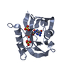
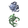
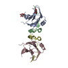
Entry Database : PDB / ID : 4c9wTitle Crystal structure of NUDT1 (MTH1) with R-crizotinib 7,8-DIHYDRO-8-OXOGUANINE TRIPHOSPHATASE Keywords / Function / homology Function Domain/homology Component
/ / / / / / / / / / / / / / / / / / / / / / / / / / / / / / / / / / / / / / / / / / / / / / / / / / / / / / / / Biological species HOMO SAPIENS (human)Method / / / Resolution : 1.65 Å Authors Elkins, J.M. / Salah, E. / Huber, K. / Superti-Furga, G. / Abdul Azeez, K.R. / Raynor, J. / Krojer, T. / von Delft, F. / Bountra, C. / Edwards, A. / Knapp, S. Journal : Nature / Year : 2014Title : Stereospecific Targeting of Mth1 by (S)-Crizotinib as an Anticancer Strategy.Authors: Huber, K.V.M. / Salah, E. / Radic, B. / Gridling, M. / Elkins, J.M. / Stukalov, A. / Jemth, A. / Gokturk, C. / Sanjiv, K. / Stromberg, K. / Pham, T. / Berglund, U.W. / Colinge, J. / Bennett, ... Authors : Huber, K.V.M. / Salah, E. / Radic, B. / Gridling, M. / Elkins, J.M. / Stukalov, A. / Jemth, A. / Gokturk, C. / Sanjiv, K. / Stromberg, K. / Pham, T. / Berglund, U.W. / Colinge, J. / Bennett, K.L. / Loizou, J.I. / Helleday, T. / Knapp, S. / Superti-Furga, G. History Deposition Oct 3, 2013 Deposition site / Processing site Revision 1.0 Apr 2, 2014 Provider / Type Revision 1.1 Apr 16, 2014 Group Revision 1.2 Jan 24, 2018 Group / Category / Item Revision 1.3 Dec 20, 2023 Group Data collection / Database references ... Data collection / Database references / Derived calculations / Other / Refinement description Category chem_comp_atom / chem_comp_bond ... chem_comp_atom / chem_comp_bond / database_2 / pdbx_database_status / pdbx_initial_refinement_model / struct_site Item _database_2.pdbx_DOI / _database_2.pdbx_database_accession ... _database_2.pdbx_DOI / _database_2.pdbx_database_accession / _pdbx_database_status.status_code_sf / _struct_site.pdbx_auth_asym_id / _struct_site.pdbx_auth_comp_id / _struct_site.pdbx_auth_seq_id
Show all Show less
 Movie
Movie Controller


 Open data
 Basic information
 Components
 Keywords
 HYDROLASE /
  Function and homology information
 Authors
 Citation
 Structure visualization
 Downloads & links
 Other downloads


 PDBj
PDBj

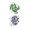
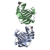
 Assembly


 Mass: 35.453 Da / Num. of mol.: 1 / Source method: obtained synthetically / Formula: Cl
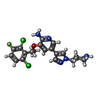
 Mass: 450.337 Da / Num. of mol.: 1 / Source method: obtained synthetically / Formula: C21H22Cl2FN5O / Comment: medication, anticancer*YM
 Mass: 96.063 Da / Num. of mol.: 5 / Source method: obtained synthetically / Formula: SO4
 Mass: 18.015 Da / Num. of mol.: 156 / Source method: isolated from a natural source / Formula: H2O
 Sample preparation
 / Beamline: I04 / Wavelength: 0.9795
  Processing