 Movie
Movie Controller


+ Open data

- Basic information
| Entry | Database: PDB / ID: 3akp | |||||||||
|---|---|---|---|---|---|---|---|---|---|---|
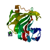
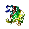
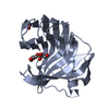
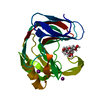
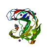
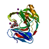
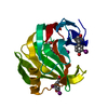
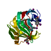
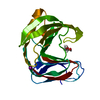
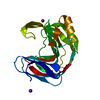
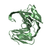
| Title | Crystal structure of xylanase from Trichoderma longibrachiatum | |||||||||
 Components Components | xylanase | |||||||||
 Keywords Keywords | HYDROLASE / xylanase | |||||||||
| Function / homology |  Function and homology informationendo-1,4-beta-xylanase activity / endo-1,4-beta-xylanase / xylan catabolic process Function and homology informationendo-1,4-beta-xylanase activity / endo-1,4-beta-xylanase / xylan catabolic processSimilarity search - Function | |||||||||
| Biological species |  Trichoderma longibrachiatum (fungus) Trichoderma longibrachiatum (fungus) | |||||||||
| Method | X-RAY DIFFRACTION / SYNCHROTRON / FOURIER SYNTHESIS / Resolution: 1.2 Å | |||||||||
 Authors Authors | Sugahara, M. / Kunishima, N. | |||||||||
 Citation Citation | Journal: Cryst.Growth Des. / Year: 2011 Title: Packing Space Expansion of Protein Crystallization Screening with Synthetic Zeolite as a Heteroepitaxic Nucleant Authors: Sugahara, M. / Kageyama-Morikawa, Y. / Kunishima, N. | |||||||||
| History |
|
- Structure visualization
| Structure viewer | Molecule: MolmilJmol/JSmol |
|---|
- Downloads & links
-Download
| PDBx/mmCIF format | 3akp.cif.gz | 92 KB | Display | PDBx/mmCIF format |
|---|---|---|---|---|
| PDB format | pdb3akp.ent.gz | 73.7 KB | Display | PDB format |
| PDBx/mmJSON format | 3akp.json.gz | Tree view | PDBx/mmJSON format | |
| Others |  Other downloads Other downloads |
-Validation report
| Arichive directory | https://data.pdbj.org/pub/pdb/validation_reports/ak/3akpftp://data.pdbj.org/pub/pdb/validation_reports/ak/3akp | HTTPS FTP |
|---|
-Related structure data


-Links
 PDBj
PDBj
- Assembly
| Deposited unit | 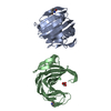
| ||||||||
|---|---|---|---|---|---|---|---|---|---|
| 1 | 
| ||||||||
| 2 | 
| ||||||||
| Unit cell |
|
-Components
| #1: Protein | Mass: 20838.436 Da / Num. of mol.: 2 Source method: isolated from a genetically manipulated source Source: (gene. exp.) Trichoderma longibrachiatum (fungus) / Production host:  Escherichia coli (E. coli) / References: UniProt: F8W669*PLUS, endo-1,4-beta-xylanase Escherichia coli (E. coli) / References: UniProt: F8W669*PLUS, endo-1,4-beta-xylanase#2: Chemical | Glycerol  Mass: 92.094 Da / Num. of mol.: 3 / Source method: obtained synthetically / Formula: C3H8O3 Mass: 92.094 Da / Num. of mol.: 3 / Source method: obtained synthetically / Formula: C3H8O3#3: Water | ChemComp-HOH / | Water Mass: 18.015 Da / Num. of mol.: 526 / Source method: isolated from a natural source / Formula: H2O Mass: 18.015 Da / Num. of mol.: 526 / Source method: isolated from a natural source / Formula: H2OSequence details | THERE IS NO DATABASE REFERENCE SEQUENCE AT PROCESSING | |
|---|
-Experimental details
-Experiment
| Experiment | Method: X-RAY DIFFRACTION / Number of used crystals: 1 |
|---|
- Sample preparation
| Crystal | Density Matthews: 2.14 Å3/Da / Density % sol: 42.45 % |
|---|---|
| Crystal grow | Temperature: 293 K / Method: oil microbatch / pH: 8.5 Details: 30% PEG 4000, 0.2M sodium acetate, pH 8.5, oil microbatch, temperature 293K |
-Data collection
| Diffraction | Mean temperature: 100 K |
|---|---|
| Diffraction source | Source: SYNCHROTRON / Site: SPring-8  / Beamline: BL26B1 / Wavelength: 0.8 Å / Beamline: BL26B1 / Wavelength: 0.8 Å |
| Detector | Type: RIGAKU RAXIS V / Detector: IMAGE PLATE / Date: Jan 28, 2010 |
| Radiation | Protocol: SINGLE WAVELENGTH / Monochromatic (M) / Laue (L): M / Scattering type: x-ray |
| Radiation wavelength | Wavelength: 0.8 Å / Relative weight: 1 |
| Reflection | Resolution: 1.2→40 Å / Num. all: 105854 / Num. obs: 105854 / % possible obs: 95.5 % / Redundancy: 3.6 % / Biso Wilson estimate: 6 Å2 / Rmerge(I) obs: 0.087 / Rsym value: 0.076 / Net I/σ(I): 6.8 |
| Reflection shell | Resolution: 1.2→1.24 Å / Redundancy: 3.6 % / Rmerge(I) obs: 0.495 / Mean I/σ(I) obs: 3 / Num. unique all: 9839 / Rsym value: 0.442 / % possible all: 89.8 |
- Processing
| Software |
| |||||||||||||||||||||||||
|---|---|---|---|---|---|---|---|---|---|---|---|---|---|---|---|---|---|---|---|---|---|---|---|---|---|---|
| Refinement | Method to determine structure: FOURIER SYNTHESIS / Resolution: 1.2→22.83 Å / Cross valid method: THROUGHOUT / σ(F): 0 / Stereochemistry target values: Engh & Huber
| |||||||||||||||||||||||||
| Displacement parameters | Biso mean: 9.1 Å2
| |||||||||||||||||||||||||
| Refine analyze |
| |||||||||||||||||||||||||
| Refinement step | Cycle: LAST / Resolution: 1.2→22.83 Å
| |||||||||||||||||||||||||
| Refine LS restraints |
| |||||||||||||||||||||||||
| LS refinement shell | Resolution: 1.2→1.24 Å / Rfactor Rfree error: 0.013
|
