 Movie
Movie Controller

[English] 日本語
 Yorodumi
- PDB-4bt9: CRYSTAL STRUCTURE OF THE PEPTIDE(PRO-PRO-GLY)3 BOUND COMPLEX OF N... -
+ Open data

- Basic information
| Entry | Database: PDB / ID: 4bt9 | ||||||
|---|---|---|---|---|---|---|---|
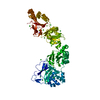
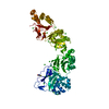
| Title | CRYSTAL STRUCTURE OF THE PEPTIDE(PRO-PRO-GLY)3 BOUND COMPLEX OF N- TERMINAL DOMAIN AND PEPTIDE SUBSTRATE BINDING DOMAIN OF PROLYL-4 HYDROXYLASE (RESIDUES 1-238) TYPE I FROM HUMAN | ||||||
 Components Components |
| ||||||
 Keywords Keywords |  OXIDOREDUCTASE / TETRATRICOPEPTIDE REPEAT MOTIF / COILED-COIL / PROLINE RICH PEPTIDE OXIDOREDUCTASE / TETRATRICOPEPTIDE REPEAT MOTIF / COILED-COIL / PROLINE RICH PEPTIDE | ||||||
| Function / homology |  Function and homology information Function and homology informationprocollagen-proline 4-dioxygenase / procollagen-proline 4-dioxygenase complex / procollagen-proline 4-dioxygenase activity / Collagen biosynthesis and modifying enzymes / L-ascorbic acid binding / collagen fibril organization / iron ion binding / endoplasmic reticulum lumen / intracellular membrane-bounded organelle / endoplasmic reticulum ...procollagen-proline 4-dioxygenase / procollagen-proline 4-dioxygenase complex / procollagen-proline 4-dioxygenase activity / Collagen biosynthesis and modifying enzymes / L-ascorbic acid binding / collagen fibril organization / iron ion binding / endoplasmic reticulum lumen / intracellular membrane-bounded organelle / endoplasmic reticulum / mitochondrion / membrane / identical protein bindingSimilarity search - Function | ||||||
| Biological species |  HOMO SAPIENS (human) HOMO SAPIENS (human)SYNTHETIC CONSTRUCT (others) | ||||||
| Method | X-RAY DIFFRACTION / SYNCHROTRON / MOLECULAR REPLACEMENT / Resolution: 1.9 Å | ||||||
 Authors Authors | Anantharajan, J. / Koski, M.K. / Wierenga, R.K. | ||||||
 Citation Citation | Journal: Structure / Year: 2013 Title: The Structural Motifs for Substrate Binding and Dimerization of the Alpha Subunit of Collagen Prolyl 4-Hydroxylase Authors: Anantharajan, J. / Koski, M.K. / Kursula, P. / Hieta, R. / Bergmann, U. / Myllyharju, J. / Wierenga, R.K. | ||||||
| History |
|
- Structure visualization
| Structure viewer | Molecule: MolmilJmol/JSmol |
|---|
- Downloads & links
-Download
| PDBx/mmCIF format | 4bt9.cif.gz | 208.6 KB | Display | PDBx/mmCIF format |
|---|---|---|---|---|
| PDB format | pdb4bt9.ent.gz | 169.5 KB | Display | PDB format |
| PDBx/mmJSON format | 4bt9.json.gz | Tree view | PDBx/mmJSON format | |
| Others |  Other downloads Other downloads |
-Validation report
| Arichive directory | https://data.pdbj.org/pub/pdb/validation_reports/bt/4bt9ftp://data.pdbj.org/pub/pdb/validation_reports/bt/4bt9 | HTTPS FTP |
|---|
-Related structure data
| Related structure data |  2yq8SC  4bt8C  4btaC  4btbC C: citing same article ( S: Starting model for refinement |
|---|---|
| Similar structure data |


-Links
 PDBj
PDBj


- Assembly
| Deposited unit | 
| ||||||||
|---|---|---|---|---|---|---|---|---|---|
| 1 |
| ||||||||
| Unit cell |
|
-Components
| #1: Protein | Procollagen-proline dioxygenase / 4-PH ALPHA-1 / PROCOLLAGEN-PROLINE\ / 2-OXOGLUTARATE-4-DIOXYGENASE SUBUNIT ALPHA-1 / PROLYL-4 ...4-PH ALPHA-1 / PROCOLLAGEN-PROLINE\ / 2-OXOGLUTARATE-4-DIOXYGENASE SUBUNIT ALPHA-1 / PROLYL-4 HYDROXYLASE TYPE I Mass: 27519.084 Da / Num. of mol.: 2 / Fragment: COLLAGEN BINDING DOMAIN, RESIDUES 18-255 Source method: isolated from a genetically manipulated source Source: (gene. exp.) HOMO SAPIENS (human) / Plasmid: PET22B / Production host:  ESCHERICHIA COLI (E. coli) / Strain (production host): BL21(DE3) ESCHERICHIA COLI (E. coli) / Strain (production host): BL21(DE3)References: UniProt: P13674, procollagen-proline 4-dioxygenase #2: Protein/peptide | ( | Mass: 771.859 Da / Num. of mol.: 1 / Source method: obtained synthetically / Source: (synth.) SYNTHETIC CONSTRUCT (others) #3: Water | ChemComp-HOH / | Water Mass: 18.015 Da / Num. of mol.: 281 / Source method: isolated from a natural source / Formula: H2O Mass: 18.015 Da / Num. of mol.: 281 / Source method: isolated from a natural source / Formula: H2O |
|---|
-Experimental details
-Experiment
| Experiment | Method: X-RAY DIFFRACTION / Number of used crystals: 2 |
|---|
- Sample preparation
| Crystal | Density Matthews: 2.33 Å3/Da / Density % sol: 47.7 % / Description: NONE |
|---|---|
| Crystal grow | pH: 6 Details: 10% PEGMME 5000, 10% DMSO, 6% MPD, 0.1 M MES, PH 6.0, 5MM SPERMINE HCL, 5MM (PRO-PRO-GLY)3 |
-Data collection
| Diffraction | Mean temperature: 100 K | |||||||||
|---|---|---|---|---|---|---|---|---|---|---|
| Diffraction source | Source: SYNCHROTRON / Site: MAX II  / Beamline: I911-3 / Wavelength: 1.0, 1.9 / Beamline: I911-3 / Wavelength: 1.0, 1.9 | |||||||||
| Detector | Type: MARRESEARCH / Detector: CCD / Date: Nov 19, 2010 | |||||||||
| Radiation | Protocol: SINGLE WAVELENGTH / Monochromatic (M) / Laue (L): M / Scattering type: x-ray | |||||||||
| Radiation wavelength |
| |||||||||
| Reflection | Resolution: 1.9→40 Å / Num. obs: 40153 / % possible obs: 99.9 % / Observed criterion σ(I): 2 / Redundancy: 19.5 % / Biso Wilson estimate: 34.76 Å2 / Rmerge(I) obs: 0.09 / Net I/σ(I): 21.9 | |||||||||
| Reflection shell | Resolution: 1.9→1.95 Å / Redundancy: 4.7 % / Rmerge(I) obs: 0.58 / Mean I/σ(I) obs: 2.4 / % possible all: 99.8 |
- Processing
| Software |
| |||||||||||||||||||||||||||||||||||||||||||||||||||||||||||||||||||||||||||||||||||||||||||||||||||||||||||||||||||||||||||||||||||||||||||||||||||||||||||||||||||||||||||||||||||||||||||||||||||||||||||||||||||||||||||||||||||||||||||||||||||||||||||||||||||||||||||||||||||
|---|---|---|---|---|---|---|---|---|---|---|---|---|---|---|---|---|---|---|---|---|---|---|---|---|---|---|---|---|---|---|---|---|---|---|---|---|---|---|---|---|---|---|---|---|---|---|---|---|---|---|---|---|---|---|---|---|---|---|---|---|---|---|---|---|---|---|---|---|---|---|---|---|---|---|---|---|---|---|---|---|---|---|---|---|---|---|---|---|---|---|---|---|---|---|---|---|---|---|---|---|---|---|---|---|---|---|---|---|---|---|---|---|---|---|---|---|---|---|---|---|---|---|---|---|---|---|---|---|---|---|---|---|---|---|---|---|---|---|---|---|---|---|---|---|---|---|---|---|---|---|---|---|---|---|---|---|---|---|---|---|---|---|---|---|---|---|---|---|---|---|---|---|---|---|---|---|---|---|---|---|---|---|---|---|---|---|---|---|---|---|---|---|---|---|---|---|---|---|---|---|---|---|---|---|---|---|---|---|---|---|---|---|---|---|---|---|---|---|---|---|---|---|---|---|---|---|---|---|---|---|---|---|---|---|---|---|---|---|---|---|---|---|---|---|---|---|---|---|---|---|---|---|---|---|---|---|---|---|---|---|---|---|---|---|---|---|---|---|---|---|---|---|---|---|---|---|
| Refinement | Method to determine structure: MOLECULAR REPLACEMENT Starting model: PDB ENTRY 2YQ8 Resolution: 1.9→58.765 Å / SU ML: 0.18 / σ(F): 1.99 / Phase error: 21.02 / Stereochemistry target values: ML
| |||||||||||||||||||||||||||||||||||||||||||||||||||||||||||||||||||||||||||||||||||||||||||||||||||||||||||||||||||||||||||||||||||||||||||||||||||||||||||||||||||||||||||||||||||||||||||||||||||||||||||||||||||||||||||||||||||||||||||||||||||||||||||||||||||||||||||||||||||
| Solvent computation | Shrinkage radii: 0.9 Å / VDW probe radii: 1.11 Å / Solvent model: FLAT BULK SOLVENT MODEL | |||||||||||||||||||||||||||||||||||||||||||||||||||||||||||||||||||||||||||||||||||||||||||||||||||||||||||||||||||||||||||||||||||||||||||||||||||||||||||||||||||||||||||||||||||||||||||||||||||||||||||||||||||||||||||||||||||||||||||||||||||||||||||||||||||||||||||||||||||
| Displacement parameters | Biso mean: 31.2 Å2 | |||||||||||||||||||||||||||||||||||||||||||||||||||||||||||||||||||||||||||||||||||||||||||||||||||||||||||||||||||||||||||||||||||||||||||||||||||||||||||||||||||||||||||||||||||||||||||||||||||||||||||||||||||||||||||||||||||||||||||||||||||||||||||||||||||||||||||||||||||
| Refinement step | Cycle: LAST / Resolution: 1.9→58.765 Å
| |||||||||||||||||||||||||||||||||||||||||||||||||||||||||||||||||||||||||||||||||||||||||||||||||||||||||||||||||||||||||||||||||||||||||||||||||||||||||||||||||||||||||||||||||||||||||||||||||||||||||||||||||||||||||||||||||||||||||||||||||||||||||||||||||||||||||||||||||||
| Refine LS restraints |
| |||||||||||||||||||||||||||||||||||||||||||||||||||||||||||||||||||||||||||||||||||||||||||||||||||||||||||||||||||||||||||||||||||||||||||||||||||||||||||||||||||||||||||||||||||||||||||||||||||||||||||||||||||||||||||||||||||||||||||||||||||||||||||||||||||||||||||||||||||
| LS refinement shell |
| |||||||||||||||||||||||||||||||||||||||||||||||||||||||||||||||||||||||||||||||||||||||||||||||||||||||||||||||||||||||||||||||||||||||||||||||||||||||||||||||||||||||||||||||||||||||||||||||||||||||||||||||||||||||||||||||||||||||||||||||||||||||||||||||||||||||||||||||||||
| Refinement TLS params. | Method: refined / Refine-ID: X-RAY DIFFRACTION
| |||||||||||||||||||||||||||||||||||||||||||||||||||||||||||||||||||||||||||||||||||||||||||||||||||||||||||||||||||||||||||||||||||||||||||||||||||||||||||||||||||||||||||||||||||||||||||||||||||||||||||||||||||||||||||||||||||||||||||||||||||||||||||||||||||||||||||||||||||
| Refinement TLS group |
|
