 Movie
Movie Controller


+ Open data

- Basic information
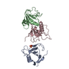
| Entry | Database: PDB / ID: 4bou | ||||||
|---|---|---|---|---|---|---|---|
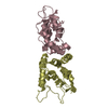
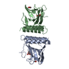
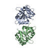
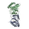
| Title | Structure of OTUD3 OTU domain | ||||||
 Components Components | OTU DOMAIN-CONTAINING PROTEIN 3 | ||||||
 Keywords Keywords |  HYDROLASE HYDROLASE | ||||||
| Function / homology |  Function and homology information Function and homology informationprotein K6-linked deubiquitination / cysteine-type deubiquitinase activity => GO:0004843 / protein K27-linked deubiquitination / protein K11-linked deubiquitination / protein K48-linked deubiquitination / protein deubiquitination / negative regulation of phosphatidylinositol 3-kinase/protein kinase B signal transduction / Regulation of PTEN stability and activity / Ovarian tumor domain proteases / ubiquitinyl hydrolase 1 ...protein K6-linked deubiquitination / cysteine-type deubiquitinase activity => GO:0004843 / protein K27-linked deubiquitination / protein K11-linked deubiquitination / protein K48-linked deubiquitination / protein deubiquitination / negative regulation of phosphatidylinositol 3-kinase/protein kinase B signal transduction / Regulation of PTEN stability and activity / Ovarian tumor domain proteases / ubiquitinyl hydrolase 1 / cysteine-type deubiquitinase activity / protein stabilization / cytosol / cytoplasmSimilarity search - Function | ||||||
| Biological species |  HOMO SAPIENS (human) HOMO SAPIENS (human) | ||||||
| Method | X-RAY DIFFRACTION / SYNCHROTRON / MOLECULAR REPLACEMENT / Resolution: 1.55 Å | ||||||
 Authors Authors | Mevissen, T.E.T. / Hospenthal, M.K. / Geurink, P.P. / Elliott, P.R. / Akutsu, M. / Arnaudo, N. / Ekkebus, R. / Kulathu, Y. / Wauer, T. / El Oualid, F. ...Mevissen, T.E.T. / Hospenthal, M.K. / Geurink, P.P. / Elliott, P.R. / Akutsu, M. / Arnaudo, N. / Ekkebus, R. / Kulathu, Y. / Wauer, T. / El Oualid, F. / Freund, S.M.V. / Ovaa, H. / Komander, D. | ||||||
 Citation Citation | Journal: Cell(Cambridge,Mass.) / Year: 2013 Title: Otu Deubiquitinases Reveal Mechanisms of Linkage Specificity and Enable Ubiquitin Chain Restriction Analysis. Authors: Mevissen, T.E.T. / Hospenthal, M.K. / Geurink, P.P. / Elliott, P.R. / Akutsu, M. / Arnaudo, N. / Ekkebus, R. / Kulathu, Y. / Wauer, T. / El Oualid, F. / Freund, S.M.V. / Ovaa, H. / Komander, D. | ||||||
| History |
|
- Structure visualization
| Structure viewer | Molecule: MolmilJmol/JSmol |
|---|
- Downloads & links
-Download
| PDBx/mmCIF format | 4bou.cif.gz | 74.9 KB | Display | PDBx/mmCIF format |
|---|---|---|---|---|
| PDB format | pdb4bou.ent.gz | 55 KB | Display | PDB format |
| PDBx/mmJSON format | 4bou.json.gz | Tree view | PDBx/mmJSON format | |
| Others |  Other downloads Other downloads |
-Validation report
| Arichive directory | https://data.pdbj.org/pub/pdb/validation_reports/bo/4bouftp://data.pdbj.org/pub/pdb/validation_reports/bo/4bou | HTTPS FTP |
|---|
-Related structure data
| Related structure data | 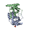 4bopC 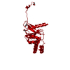 4boqC  4bosC  4bozC 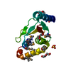 3pfyS C: citing same article ( S: Starting model for refinement |
|---|---|
| Similar structure data |


-Links
 PDBj
PDBj- Assembly
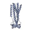
| Deposited unit | 
| ||||||||
|---|---|---|---|---|---|---|---|---|---|
| 1 | 
| ||||||||
| Unit cell |
| ||||||||
| Components on special symmetry positions |
|
-Components
| #1: Protein | Mass: 18353.455 Da / Num. of mol.: 1 / Fragment: RESIDUES 52-209 Source method: isolated from a genetically manipulated source Source: (gene. exp.) HOMO SAPIENS (human) / Plasmid: POPINK / Production host:  ESCHERICHIA COLI (E. coli) / Strain (production host): BL21(DE3) / Variant (production host): ROSETTA PLACI / References: UniProt: Q5T2D3, ubiquitinyl hydrolase 1 ESCHERICHIA COLI (E. coli) / Strain (production host): BL21(DE3) / Variant (production host): ROSETTA PLACI / References: UniProt: Q5T2D3, ubiquitinyl hydrolase 1 |
|---|---|
| #2: Water | ChemComp-HOH / Water Mass: 18.015 Da / Num. of mol.: 113 / Source method: isolated from a natural source / Formula: H2O Mass: 18.015 Da / Num. of mol.: 113 / Source method: isolated from a natural source / Formula: H2O |
| Sequence details | CONTAINS ADDITIONAL |
-Experimental details
-Experiment
| Experiment | Method: X-RAY DIFFRACTION / Number of used crystals: 1 |
|---|
- Sample preparation
| Crystal | Density Matthews: 2.26 Å3/Da / Density % sol: 45.6 % / Description: NONE |
|---|---|
| Crystal grow | Details: 10% PEG 4K, 0.1 M KCL, 0.01 M MGCL2, 50 MM MES PH 6.0 |
-Data collection
| Diffraction | Mean temperature: 287 K |
|---|---|
| Diffraction source | Source: SYNCHROTRON / Site: ESRF  / Beamline: ID29 / Wavelength: 0.97627 / Beamline: ID29 / Wavelength: 0.97627 |
| Detector | Type: ADSC CCD / Detector: CCD / Details: MIRROR |
| Radiation | Monochromator: NI FILTER / Protocol: SINGLE WAVELENGTH / Monochromatic (M) / Laue (L): M / Scattering type: x-ray |
| Radiation wavelength | Wavelength: 0.97627 Å / Relative weight: 1 |
| Reflection | Resolution: 1.55→42.21 Å / Num. obs: 23429 / % possible obs: 98.9 % / Observed criterion σ(I): 2 / Redundancy: 3.9 % / Biso Wilson estimate: 23.66 Å2 / Rmerge(I) obs: 0.04 / Net I/σ(I): 14.9 |
| Reflection shell | Resolution: 1.55→1.63 Å / Redundancy: 3.9 % / Rmerge(I) obs: 0.48 / Mean I/σ(I) obs: 2.1 / % possible all: 99.1 |
- Processing
| Software |
| ||||||||||||||||||||||||||||||||||||||||||||||||||||||||||||||||||||||
|---|---|---|---|---|---|---|---|---|---|---|---|---|---|---|---|---|---|---|---|---|---|---|---|---|---|---|---|---|---|---|---|---|---|---|---|---|---|---|---|---|---|---|---|---|---|---|---|---|---|---|---|---|---|---|---|---|---|---|---|---|---|---|---|---|---|---|---|---|---|---|---|
| Refinement | Method to determine structure: MOLECULAR REPLACEMENT Starting model: PDB ENTRY 3PFY Resolution: 1.55→31.347 Å / SU ML: 0.2 / σ(F): 1.35 / Phase error: 24.01 / Stereochemistry target values: ML
| ||||||||||||||||||||||||||||||||||||||||||||||||||||||||||||||||||||||
| Solvent computation | Shrinkage radii: 0.9 Å / VDW probe radii: 1.11 Å / Solvent model: FLAT BULK SOLVENT MODEL | ||||||||||||||||||||||||||||||||||||||||||||||||||||||||||||||||||||||
| Refinement step | Cycle: LAST / Resolution: 1.55→31.347 Å
| ||||||||||||||||||||||||||||||||||||||||||||||||||||||||||||||||||||||
| Refine LS restraints |
| ||||||||||||||||||||||||||||||||||||||||||||||||||||||||||||||||||||||
| LS refinement shell |
|
