 Movie
Movie Controller

[English] 日本語
 Yorodumi
- PDB-6hqz: Crystal structure of the type III effector protein AvrRpt2 from E... -
+ Open data

- Basic information
| Entry | Database: PDB / ID: 6hqz | ||||||
|---|---|---|---|---|---|---|---|
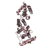
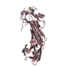
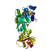
| Title | Crystal structure of the type III effector protein AvrRpt2 from Erwinia amylovora, a C70 family cysteine protease | ||||||
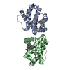
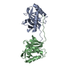
 Components Components | AvrRpt2 | ||||||
 Keywords Keywords |  HYDROLASE / Avirulence / resistance / plant pathogen / cysteine protease / effector / T3ss HYDROLASE / Avirulence / resistance / plant pathogen / cysteine protease / effector / T3ss | ||||||
| Function / homology | Peptidase C70, AvrRpt2 / Papain-like cysteine protease AvrRpt2 / AvrRpt2 Function and homology information Function and homology information | ||||||
| Biological species |  Erwinia amylovora (bacteria) Erwinia amylovora (bacteria) | ||||||
| Method | X-RAY DIFFRACTION / SYNCHROTRON / MAD / Resolution: 1.8 Å | ||||||
 Authors Authors | Bartho, J.D. / Demitri, N. / Wuerges, J. / Benini, S. | ||||||
 Citation Citation | Journal: J.Struct.Biol. / Year: 2019 Title: The structure of Erwinia amylovora AvrRpt2 provides insight into protein maturation and induced resistance to fire blight by Malus × robusta 5. Authors: Bartho, J.D. / Demitri, N. / Bellini, D. / Flachowsky, H. / Peil, A. / Walsh, M.A. / Benini, S. | ||||||
| History |
|
- Structure visualization
| Structure viewer | Molecule: MolmilJmol/JSmol |
|---|
- Downloads & links
-Download
| PDBx/mmCIF format | 6hqz.cif.gz | 133.1 KB | Display | PDBx/mmCIF format |
|---|---|---|---|---|
| PDB format | pdb6hqz.ent.gz | 109.2 KB | Display | PDB format |
| PDBx/mmJSON format | 6hqz.json.gz | Tree view | PDBx/mmJSON format | |
| Others |  Other downloads Other downloads |
-Validation report
| Arichive directory | https://data.pdbj.org/pub/pdb/validation_reports/hq/6hqzftp://data.pdbj.org/pub/pdb/validation_reports/hq/6hqz | HTTPS FTP |
|---|
-Related structure data
| Similar structure data |
|---|


-Links
 PDBj
PDBj
- Assembly
| Deposited unit | 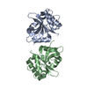
| ||||||||
|---|---|---|---|---|---|---|---|---|---|
| 1 |
| ||||||||
| Unit cell |
|
-Components
| #1: Protein | Mass: 17494.756 Da / Num. of mol.: 2 Source method: isolated from a genetically manipulated source Details: In this construct the protein sequence starts from G70, N69 is a result of the cloning procedure and of subsequent tag cleavage. we report the structure of AvrRpt2_70-222. A S-S bridge is ...Details: In this construct the protein sequence starts from G70, N69 is a result of the cloning procedure and of subsequent tag cleavage. we report the structure of AvrRpt2_70-222. A S-S bridge is formed between C156 of chain A and C156 of chain B Source: (gene. exp.) Erwinia amylovora (bacteria) / Production host: Escherichia coli BL21(DE3) (bacteria) / References: UniProt: A0A2U7NR52#2: Water | ChemComp-HOH / | Water Mass: 18.015 Da / Num. of mol.: 91 / Source method: isolated from a natural source / Formula: H2O Mass: 18.015 Da / Num. of mol.: 91 / Source method: isolated from a natural source / Formula: H2O |
|---|
-Experimental details
-Experiment
| Experiment | Method: X-RAY DIFFRACTION / Number of used crystals: 1 |
|---|
- Sample preparation
| Crystal | Density Matthews: 2.32 Å3/Da / Density % sol: 47 % |
|---|---|
| Crystal grow | Temperature: 293 K / Method: microbatch / pH: 9 Details: 100 mM TRIS-HCL pH 9.0 10% glycerol, 100 mM sodium cacodylate, 15% PEG 6K Temp details: vibration free incubator |
-Data collection
| Diffraction | Mean temperature: 100 K |
|---|---|
| Diffraction source | Source: SYNCHROTRON / Site: ELETTRA  / Beamline: 5.2R / Wavelength: 1.064 Å / Beamline: 5.2R / Wavelength: 1.064 Å |
| Detector | Type: DECTRIS PILATUS 2M / Detector: PIXEL / Date: Sep 21, 2014 |
| Radiation | Protocol: SINGLE WAVELENGTH / Monochromatic (M) / Laue (L): M / Scattering type: x-ray |
| Radiation wavelength | Wavelength: 1.064 Å / Relative weight: 1 |
| Reflection | Resolution: 1.8→51.32 Å / Num. obs: 28980 / % possible obs: 99.02 % / Redundancy: 2.8 % / Rrim(I) all: 0.054 / Net I/σ(I): 13.4 |
| Reflection shell | Resolution: 1.8→1.85 Å / Num. unique obs: 2004 / Rrim(I) all: 0.664 / % possible all: 94.76 |
- Processing
| Software |
| ||||||||||||||||||||||||||||||||||||||||||||||||||||||||||||||||||||||||||||||||||||||||||||||||||||||||||||||||||||||||||||||||||||||||||||||||||||||||||||||||||||||||||||||||||||||
|---|---|---|---|---|---|---|---|---|---|---|---|---|---|---|---|---|---|---|---|---|---|---|---|---|---|---|---|---|---|---|---|---|---|---|---|---|---|---|---|---|---|---|---|---|---|---|---|---|---|---|---|---|---|---|---|---|---|---|---|---|---|---|---|---|---|---|---|---|---|---|---|---|---|---|---|---|---|---|---|---|---|---|---|---|---|---|---|---|---|---|---|---|---|---|---|---|---|---|---|---|---|---|---|---|---|---|---|---|---|---|---|---|---|---|---|---|---|---|---|---|---|---|---|---|---|---|---|---|---|---|---|---|---|---|---|---|---|---|---|---|---|---|---|---|---|---|---|---|---|---|---|---|---|---|---|---|---|---|---|---|---|---|---|---|---|---|---|---|---|---|---|---|---|---|---|---|---|---|---|---|---|---|---|
| Refinement | Method to determine structure: MAD / Resolution: 1.8→51.32 Å / Cor.coef. Fo:Fc: 0.956 / Cor.coef. Fo:Fc free: 0.951 / SU B: 7.938 / SU ML: 0.114 / Cross valid method: THROUGHOUT / ESU R: 0.142 / ESU R Free: 0.127 / Details: HYDROGENS HAVE BEEN ADDED IN THE RIDING POSITIONS
| ||||||||||||||||||||||||||||||||||||||||||||||||||||||||||||||||||||||||||||||||||||||||||||||||||||||||||||||||||||||||||||||||||||||||||||||||||||||||||||||||||||||||||||||||||||||
| Solvent computation | Ion probe radii: 0.8 Å / Shrinkage radii: 0.8 Å / VDW probe radii: 1.2 Å | ||||||||||||||||||||||||||||||||||||||||||||||||||||||||||||||||||||||||||||||||||||||||||||||||||||||||||||||||||||||||||||||||||||||||||||||||||||||||||||||||||||||||||||||||||||||
| Displacement parameters | Biso mean: 39.225 Å2
| ||||||||||||||||||||||||||||||||||||||||||||||||||||||||||||||||||||||||||||||||||||||||||||||||||||||||||||||||||||||||||||||||||||||||||||||||||||||||||||||||||||||||||||||||||||||
| Refinement step | Cycle: 1 / Resolution: 1.8→51.32 Å
| ||||||||||||||||||||||||||||||||||||||||||||||||||||||||||||||||||||||||||||||||||||||||||||||||||||||||||||||||||||||||||||||||||||||||||||||||||||||||||||||||||||||||||||||||||||||
| Refine LS restraints |
|