 Movie
Movie Controller


+ Open data

- Basic information
| Entry | Database: PDB / ID: 4bkg | ||||||
|---|---|---|---|---|---|---|---|
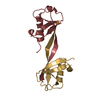
| Title | crystal structure of human diSUMO-2 | ||||||
 Components Components | SMALL UBIQUITIN-RELATED MODIFIER 2 | ||||||
 Keywords Keywords |  PROTEIN BINDING PROTEIN BINDING | ||||||
| Function / homology |  Function and homology information Function and homology informationSUMO is proteolytically processed / SUMO is conjugated to E1 (UBA2:SAE1) / SUMO is transferred from E1 to E2 (UBE2I, UBC9) / Vitamin D (calciferol) metabolism / SUMOylation of SUMOylation proteins / SUMOylation of RNA binding proteins / SUMO transferase activity / ubiquitin-like protein ligase binding / SUMOylation of DNA replication proteins / protein sumoylation ...SUMO is proteolytically processed / SUMO is conjugated to E1 (UBA2:SAE1) / SUMO is transferred from E1 to E2 (UBE2I, UBC9) / Vitamin D (calciferol) metabolism / SUMOylation of SUMOylation proteins / SUMOylation of RNA binding proteins / SUMO transferase activity / ubiquitin-like protein ligase binding / SUMOylation of DNA replication proteins / protein sumoylation / SUMOylation of transcription factors / SUMOylation of DNA damage response and repair proteins / SUMOylation of chromatin organization proteins / SUMOylation of transcription cofactors / SUMOylation of intracellular receptors / PML body / Formation of Incision Complex in GG-NER / protein tag activity / positive regulation of proteasomal ubiquitin-dependent protein catabolic process / Processing of DNA double-strand break ends / ubiquitin protein ligase binding / positive regulation of transcription by RNA polymerase II / RNA binding / nucleoplasm / nucleusSimilarity search - Function | ||||||
| Biological species |  HOMO SAPIENS (human) HOMO SAPIENS (human) | ||||||
| Method | X-RAY DIFFRACTION / MOLECULAR REPLACEMENT / Resolution: 2.11 Å | ||||||
 Authors Authors | Keusekotten, K. / Bade, V.N. / Meyer-Teschendorf, K. / Sriramachandran, A. / Fischer-Schrader, K. / Krause, A. / Horst, C. / Hofmann, K. / Dohmen, R.J. / Praefcke, G.J.K. | ||||||
 Citation Citation | Journal: Biochem.J. / Year: 2014 Title: Multivalent Interactions of the Sumo-Interaction Motifs in the Ring-Finger Protein 4 (Rnf4) Determine the Specificity for Chains of the Small Ubiquitin-Related Modifier (Sumo). Authors: Keusekotten, K. / Bade, V.N. / Meyer-Teschendorf, K. / Sriramachandran, A.M. / Fischer-Schrader, K. / Krause, A. / Horst, C. / Schwarz, G. / Hofmann, K. / Dohmen, R.J. / Praefcke, G.J. | ||||||
| History |
|
- Structure visualization
| Structure viewer | Molecule: MolmilJmol/JSmol |
|---|
- Downloads & links
-Download
| PDBx/mmCIF format | 4bkg.cif.gz | 29.8 KB | Display | PDBx/mmCIF format |
|---|---|---|---|---|
| PDB format | pdb4bkg.ent.gz | 18.3 KB | Display | PDB format |
| PDBx/mmJSON format | 4bkg.json.gz | Tree view | PDBx/mmJSON format | |
| Others |  Other downloads Other downloads |
-Validation report
| Arichive directory | https://data.pdbj.org/pub/pdb/validation_reports/bk/4bkgftp://data.pdbj.org/pub/pdb/validation_reports/bk/4bkg | HTTPS FTP |
|---|
-Related structure data
| Related structure data |  1wm3S S: Starting model for refinement |
|---|---|
| Similar structure data |


-Links
 PDBj
PDBj


- Assembly
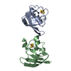
| Deposited unit | 
| ||||||||
|---|---|---|---|---|---|---|---|---|---|
| 1 | 
| ||||||||
| Unit cell |
| ||||||||
| Details | THE PROTEIN CHAIN IS SEGMENTED INTO DIFFERENT ASUS VIA A CRYSTALLOGRAPHIC SYMMETRY AXIS. THUS THE DIMERIC ASSEMBLY IS ACTUALLY A MONOMER IN WHICH THE HALVES ARE RELATED BY SYMMETRY AND LINKED VIA AN UNOBSERVED LINKER |
-Components
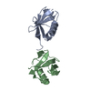
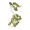
| #1: Protein | Mass: 19185.377 Da / Num. of mol.: 1 / Fragment: SUMO-2DELTAN11, RESIDUES 12-93 Source method: isolated from a genetically manipulated source Details: LINEAR FUSION-PROTEIN OF 2 SUMO2-DELTAN11 FRAGMENTS, WHICH ARE PRESENT IN DIFFERENT ASU, RELATED VIA A CRYSTALLOGRAPHIC SYMMETRY AXIS Source: (gene. exp.) HOMO SAPIENS (human) / Plasmid: PGEX-4T2 / Production host:  ESCHERICHIA COLI (E. coli) / References: UniProt: P61956 ESCHERICHIA COLI (E. coli) / References: UniProt: P61956 |
|---|---|
| #2: Water | ChemComp-HOH / Water Mass: 18.015 Da / Num. of mol.: 27 / Source method: isolated from a natural source / Formula: H2O Mass: 18.015 Da / Num. of mol.: 27 / Source method: isolated from a natural source / Formula: H2O |
-Experimental details
-Experiment
| Experiment | Method: X-RAY DIFFRACTION / Number of used crystals: 1 |
|---|
- Sample preparation
| Crystal | Density Matthews: 1.89 Å3/Da / Density % sol: 34.91 % / Description: NONE |
|---|---|
| Crystal grow | pH: 8 Details: 0.1 M TRIS/HCL PH 8.0, 28% PEG 350MME,0.05% DIOXANE |
-Data collection
| Diffraction | Mean temperature: 100 K |
|---|---|
| Diffraction source | Source: SEALED TUBE / Type: OXFORD DIFFRACTION NOVA / Wavelength: 1.5406 / Wavelength: 1.5406 Å |
| Detector | Type: OXFORD DIFFRACTION / Detector: CCD / Date: Jan 24, 2013 / Details: NOVA |
| Radiation | Protocol: SINGLE WAVELENGTH / Monochromatic (M) / Laue (L): M / Scattering type: x-ray |
| Radiation wavelength | Wavelength: 1.5406 Å / Relative weight: 1 |
| Reflection | Resolution: 2.1→23.24 Å / Num. obs: 3842 / % possible obs: 94.8 % / Observed criterion σ(I): 2 / Redundancy: 2.8 % / Rmerge(I) obs: 0.06 / Net I/σ(I): 12.5 |
| Reflection shell | Resolution: 2.1→2.17 Å / Redundancy: 1.4 % / Rmerge(I) obs: 0.44 / Mean I/σ(I) obs: 1.9 / % possible all: 73.9 |
- Processing
| Software |
| ||||||||||||||||||||||||||||||||||||||||||||||||||||||||||||||||||||||||||||||||||||||||||||||||||||||||||||||||||||||||||||||||||||||||||||||||||||||||||||||||||||||||||||||||||||||
|---|---|---|---|---|---|---|---|---|---|---|---|---|---|---|---|---|---|---|---|---|---|---|---|---|---|---|---|---|---|---|---|---|---|---|---|---|---|---|---|---|---|---|---|---|---|---|---|---|---|---|---|---|---|---|---|---|---|---|---|---|---|---|---|---|---|---|---|---|---|---|---|---|---|---|---|---|---|---|---|---|---|---|---|---|---|---|---|---|---|---|---|---|---|---|---|---|---|---|---|---|---|---|---|---|---|---|---|---|---|---|---|---|---|---|---|---|---|---|---|---|---|---|---|---|---|---|---|---|---|---|---|---|---|---|---|---|---|---|---|---|---|---|---|---|---|---|---|---|---|---|---|---|---|---|---|---|---|---|---|---|---|---|---|---|---|---|---|---|---|---|---|---|---|---|---|---|---|---|---|---|---|---|---|
| Refinement | Method to determine structure: MOLECULAR REPLACEMENT Starting model: PDB ENTRY 1WM3 Resolution: 2.11→19.76 Å / Cor.coef. Fo:Fc: 0.951 / Cor.coef. Fo:Fc free: 0.918 / SU B: 5.426 / SU ML: 0.14 / Cross valid method: THROUGHOUT / ESU R: 0.287 / ESU R Free: 0.208 / Stereochemistry target values: MAXIMUM LIKELIHOOD Details: HYDROGENS HAVE BEEN ADDED IN THE RIDING POSITIONS. RESIDUES 17-89 MAKE UP THE CONTENT OF THE ASU. RESIDUES 101-173 ARE IDENTICAL TO RESIDUES 17-89, AND SEGMENTED INTO DIFFERENT ASU VIA A ...Details: HYDROGENS HAVE BEEN ADDED IN THE RIDING POSITIONS. RESIDUES 17-89 MAKE UP THE CONTENT OF THE ASU. RESIDUES 101-173 ARE IDENTICAL TO RESIDUES 17-89, AND SEGMENTED INTO DIFFERENT ASU VIA A CRYSTALLOGRAPHIC SYMMETRY AXIS. THE GAP OF APPROX. 14 A BETWEEN THE C-TERMINUS OF A MOLECULE IN A ASU TO THE N-TERMINUS OF A SYMMETRY-RELATED MOLECULE IN THE ADJACENT ASU CAN BE FILLED BY 11 RESIDUES FROM THE LINKER (90-100) WHICH ARE DELOCALIZED. THE TERMINAL RESIDUES 10-16 AND 174-177 ARE FLEXIBLE AND NOT OBSERVED IN THE CRYSTAL STRUCTURE.
| ||||||||||||||||||||||||||||||||||||||||||||||||||||||||||||||||||||||||||||||||||||||||||||||||||||||||||||||||||||||||||||||||||||||||||||||||||||||||||||||||||||||||||||||||||||||
| Solvent computation | Ion probe radii: 0.8 Å / Shrinkage radii: 0.8 Å / VDW probe radii: 1.2 Å / Solvent model: MASK | ||||||||||||||||||||||||||||||||||||||||||||||||||||||||||||||||||||||||||||||||||||||||||||||||||||||||||||||||||||||||||||||||||||||||||||||||||||||||||||||||||||||||||||||||||||||
| Displacement parameters | Biso mean: 27.678 Å2
| ||||||||||||||||||||||||||||||||||||||||||||||||||||||||||||||||||||||||||||||||||||||||||||||||||||||||||||||||||||||||||||||||||||||||||||||||||||||||||||||||||||||||||||||||||||||
| Refinement step | Cycle: LAST / Resolution: 2.11→19.76 Å
| ||||||||||||||||||||||||||||||||||||||||||||||||||||||||||||||||||||||||||||||||||||||||||||||||||||||||||||||||||||||||||||||||||||||||||||||||||||||||||||||||||||||||||||||||||||||
| Refine LS restraints |
|