 Movie
Movie Controller


+ Open data

- Basic information
| Entry | Database: PDB / ID: 1lfm | ||||||
|---|---|---|---|---|---|---|---|
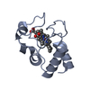
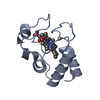
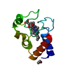
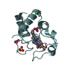
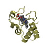
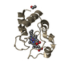
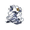
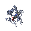
| Title | CRYSTAL STRUCTURE OF COBALT(III)-SUBSTITUTED CYTOCHROME C (TUNA) | ||||||
 Components Components | CYTOCHROME C | ||||||
 Keywords Keywords | ELECTRON TRANSPORT / CYTOCHROME C / Folding / Intermediates | ||||||
| Function / homology |  Function and homology informationrespirasome / mitochondrial intermembrane space / electron transfer activity / heme binding / metal ion binding Function and homology informationrespirasome / mitochondrial intermembrane space / electron transfer activity / heme binding / metal ion bindingSimilarity search - Function | ||||||
| Biological species |  Thunnus thynnus (Atlantic bluefin tuna) Thunnus thynnus (Atlantic bluefin tuna) | ||||||
| Method | X-RAY DIFFRACTION / rigid body refinement / Resolution: 1.5 Å | ||||||
 Authors Authors | Tezcan, F.A. / Findley, W.M. / Crane, B.R. / Ross, S.A. / Lyubovitsky, J.G. / Gray, H.B. / Winkler, J.R. | ||||||
 Citation Citation | Journal: Proc.Natl.Acad.Sci.USA / Year: 2002 Title: Using deeply trapped intermediates to map the cytochrome c folding landscape. Authors: Tezcan, F.A. / Findley, W.M. / Crane, B.R. / Ross, S.A. / Lyubovitsky, J.G. / Gray, H.B. / Winkler, J.R. | ||||||
| History |
|
- Structure visualization
| Structure viewer | Molecule: MolmilJmol/JSmol |
|---|
- Downloads & links
-Download
| PDBx/mmCIF format | 1lfm.cif.gz | 64.1 KB | Display | PDBx/mmCIF format |
|---|---|---|---|---|
| PDB format | pdb1lfm.ent.gz | 45.8 KB | Display | PDB format |
| PDBx/mmJSON format | 1lfm.json.gz | Tree view | PDBx/mmJSON format | |
| Others |  Other downloads Other downloads |
-Validation report
| Arichive directory | https://data.pdbj.org/pub/pdb/validation_reports/lf/1lfmftp://data.pdbj.org/pub/pdb/validation_reports/lf/1lfm | HTTPS FTP |
|---|
-Related structure data
| Related structure data | 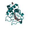 3cytS S: Starting model for refinement |
|---|---|
| Similar structure data |


-Links
 PDBj
PDBj


- Assembly
| Deposited unit | 
| ||||||||||
|---|---|---|---|---|---|---|---|---|---|---|---|
| 1 | 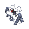
| ||||||||||
| 2 | 
| ||||||||||
| Unit cell |
|
-Components
| #1: Protein | Mass: 11390.076 Da / Num. of mol.: 2 / Source method: isolated from a natural source / Source: (natural) Thunnus thynnus (Atlantic bluefin tuna) / Organelle: mitochondriaMitochondrion / References: UniProt: P81459#2: Chemical |   Mass: 619.575 Da / Num. of mol.: 2 / Source method: obtained synthetically / Formula: C34H32CoN4O4 Mass: 619.575 Da / Num. of mol.: 2 / Source method: obtained synthetically / Formula: C34H32CoN4O4#3: Water | ChemComp-HOH / | Water Mass: 18.015 Da / Num. of mol.: 374 / Source method: isolated from a natural source / Formula: H2O Mass: 18.015 Da / Num. of mol.: 374 / Source method: isolated from a natural source / Formula: H2O |
|---|
-Experimental details
-Experiment
| Experiment | Method: X-RAY DIFFRACTION / Number of used crystals: 1 |
|---|
- Sample preparation
| Crystal | Density Matthews: 2.15 Å3/Da / Density % sol: 42.7 % | |||||||||||||||||||||||||||||||||||
|---|---|---|---|---|---|---|---|---|---|---|---|---|---|---|---|---|---|---|---|---|---|---|---|---|---|---|---|---|---|---|---|---|---|---|---|---|
| Crystal grow | Temperature: 298 K / Method: vapor diffusion, hanging drop / pH: 6.4 Details: (NH4)2SO4, NaCl, Na3PO4, pH 6.4, VAPOR DIFFUSION, HANGING DROP at 298K | |||||||||||||||||||||||||||||||||||
| Crystal grow | *PLUS pH: 6.5 | |||||||||||||||||||||||||||||||||||
| Components of the solutions | *PLUS
|
-Data collection
| Diffraction | Mean temperature: 100 K |
|---|---|
| Diffraction source | Source: ROTATING ANODE / Type: RIGAKU RU200 / Wavelength: 1.5418 Å |
| Detector | Type: RIGAKU RAXIS IV / Detector: IMAGE PLATE / Date: Sep 25, 1999 / Details: MIRRORS |
| Radiation | Protocol: SINGLE WAVELENGTH / Monochromatic (M) / Laue (L): M / Scattering type: x-ray |
| Radiation wavelength | Wavelength: 1.5418 Å / Relative weight: 1 |
| Reflection | Resolution: 1.5→15.69 Å / Num. all: 31317 / Num. obs: 27926 / % possible obs: 89.2 % / Observed criterion σ(I): -3 / Redundancy: 2.5 % / Biso Wilson estimate: 18.2 Å2 / Rsym value: 0.073 / Net I/σ(I): 17.3 |
| Reflection shell | Resolution: 1.5→1.55 Å / Redundancy: 2.5 % / Mean I/σ(I) obs: 3.2 / Num. unique all: 2738 / Rsym value: 0.242 / % possible all: 88.4 |
| Reflection | *PLUS Highest resolution: 1.5 Å / Rmerge(I) obs: 0.073 |
- Processing
| Software |
| |||||||||||||||||||||||||
|---|---|---|---|---|---|---|---|---|---|---|---|---|---|---|---|---|---|---|---|---|---|---|---|---|---|---|
| Refinement | Method to determine structure: rigid body refinement Starting model: 3CYT Resolution: 1.5→15.69 Å / Cross valid method: THROUGHOUT / σ(F): 0 / Stereochemistry target values: Engh & Huber
| |||||||||||||||||||||||||
| Refine analyze |
| |||||||||||||||||||||||||
| Refinement step | Cycle: LAST / Resolution: 1.5→15.69 Å
| |||||||||||||||||||||||||
| Refine LS restraints |
| |||||||||||||||||||||||||
| LS refinement shell | Resolution: 1.5→1.55 Å / Rfactor Rfree error: 0.024
| |||||||||||||||||||||||||
| Refinement | *PLUS Highest resolution: 1.5 Å / Rfactor obs: 0.183 / Rfactor Rfree: 0.22 / Rfactor Rwork: 0.183 | |||||||||||||||||||||||||
| Solvent computation | *PLUS | |||||||||||||||||||||||||
| Displacement parameters | *PLUS | |||||||||||||||||||||||||
| Refine LS restraints | *PLUS
| |||||||||||||||||||||||||
| LS refinement shell | *PLUS Rfactor Rfree: 0.366 / Rfactor Rwork: 0.361 |
