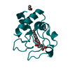
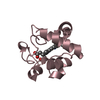
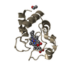
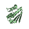
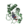
HETEROGEN The residue 1104 of HEM and residue 1105 of ZNH are alternate conformers with occupancy ...HETEROGEN The residue 1104 of HEM and residue 1105 of ZNH are alternate conformers with occupancy of 0.39 and 0.61, respectively. In similar residue 1106 of HEM and residue 1107 of ZNH are alternate conformers with occupancy 0.39 and 0.61, respectively.
Resolution: 2→19.64 Å / Rfactor Rfree error: 0.008 / Data cutoff high absF: 381490.79 / Data cutoff low absF: 0 / Isotropic thermal model: RESTRAINED / σ(F): 0 / σ(I): 0 / Stereochemistry target values: Engh & Huber Details: Crystals contained a mixture of Zn- and Fe- porphyrins with occupancies determined from anomalous scattering measurements. Residues 1104 and 1105 and residues 1105 and 1106 were refined as ...Details: Crystals contained a mixture of Zn- and Fe- porphyrins with occupancies determined from anomalous scattering measurements. Residues 1104 and 1105 and residues 1105 and 1106 were refined as non-interacting porphyrins, reflecting the mixture of Zn and Fe cytc in the crystal lattice. No stereochemical restraints were placed on ZN or Fe interactions.
In the structure databanks used in Yorodumi, some data are registered as the other names, "COVID-19 virus" and "2019-nCoV". Here are the details of the virus and the list of structure data.
Jan 31, 2019. EMDB accession codes are about to change! (news from PDBe EMDB page)
EMDB accession codes are about to change! (news from PDBe EMDB page)
The allocation of 4 digits for EMDB accession codes will soon come to an end. Whilst these codes will remain in use, new EMDB accession codes will include an additional digit and will expand incrementally as the available range of codes is exhausted. The current 4-digit format prefixed with “EMD-” (i.e. EMD-XXXX) will advance to a 5-digit format (i.e. EMD-XXXXX), and so on. It is currently estimated that the 4-digit codes will be depleted around Spring 2019, at which point the 5-digit format will come into force.
The EM Navigator/Yorodumi systems omit the EMD- prefix.
Related info.:Q: What is EMD? / ID/Accession-code notation in Yorodumi/EM Navigator
Yorodumi is a browser for structure data from EMDB, PDB, SASBDB, etc.
This page is also the successor to EM Navigator detail page, and also detail information page/front-end page for Omokage search.
The word "yorodu" (or yorozu) is an old Japanese word meaning "ten thousand". "mi" (miru) is to see.
Related info.:EMDB / PDB / SASBDB / Comparison of 3 databanks / Yorodumi Search / Aug 31, 2016. New EM Navigator & Yorodumi / Yorodumi Papers / Jmol/JSmol / Function and homology information / Changes in new EM Navigator and Yorodumi
 Movie
Movie Controller


 Open data
 Basic information
 Components
 Keywords
 Function and homology information
 Authors
 Citation
 Structure visualization
 Downloads & links
 Other downloads
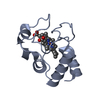


 PDBj
PDBj


 Assembly


 Mass: 618.503 Da / Num. of mol.: 2 / Source method: obtained synthetically / Formula: C34H34FeN4O4
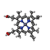
 Mass: 626.051 Da / Num. of mol.: 2 / Source method: obtained synthetically / Formula: C34H32N4O4Zn
 Mass: 18.015 Da / Num. of mol.: 370 / Source method: isolated from a natural source / Formula: H2O
 Sample preparation
 / Beamline: BL9-2 / Wavelength: 1.282 Å
 Processing