 Movie
Movie Controller


+ Open data

- Basic information
| Entry | Database: PDB / ID: 4ag0 | ||||||
|---|---|---|---|---|---|---|---|
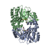
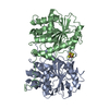
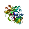
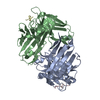
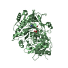
| Title | Crystal structure of FimX EAL domain | ||||||
 Components Components | FIMX | ||||||
 Keywords Keywords |  HYDROLASE / PHOSPHODIESTERASE / C-DIGMP / BIOFILM HYDROLASE / PHOSPHODIESTERASE / C-DIGMP / BIOFILM | ||||||
| Function / homology |  Function and homology information Function and homology informationphosphorelay signal transduction system / nucleotide binding / regulation of DNA-templated transcription / identical protein bindingSimilarity search - Function | ||||||
| Biological species |   PSEUDOMONAS AERUGINOSA (bacteria) PSEUDOMONAS AERUGINOSA (bacteria) | ||||||
| Method | X-RAY DIFFRACTION / SYNCHROTRON / MOLECULAR REPLACEMENT / Resolution: 2.304 Å | ||||||
 Authors Authors | Robert-Paganin, J. / Nonin-Lecomte, S. / Rety, S. | ||||||
 Citation Citation | Journal: Plos One / Year: 2012 Title: Crystal Structure of an Eal Domain in Complex with Reaction Product 5'-Pgpg Authors: Robert-Paganin, J. / Nonin-Lecomte, S. / Rety, S. | ||||||
| History |
| ||||||
| Remark 700 | SHEET DETERMINATION METHOD: DSSP THE SHEETS PRESENTED AS "BA" IN EACH CHAIN ON SHEET RECORDS BELOW ... SHEET DETERMINATION METHOD: DSSP THE SHEETS PRESENTED AS "BA" IN EACH CHAIN ON SHEET RECORDS BELOW IS ACTUALLY AN 8-STRANDED BARREL THIS IS REPRESENTED BY A 9-STRANDED SHEET IN WHICH THE FIRST AND LAST STRANDS ARE IDENTICAL. |
- Structure visualization
| Structure viewer | Molecule: MolmilJmol/JSmol |
|---|
- Downloads & links
-Download
| PDBx/mmCIF format | 4ag0.cif.gz | 112.7 KB | Display | PDBx/mmCIF format |
|---|---|---|---|---|
| PDB format | pdb4ag0.ent.gz | 86.8 KB | Display | PDB format |
| PDBx/mmJSON format | 4ag0.json.gz | Tree view | PDBx/mmJSON format | |
| Others |  Other downloads Other downloads |
-Validation report
| Arichive directory | https://data.pdbj.org/pub/pdb/validation_reports/ag/4ag0ftp://data.pdbj.org/pub/pdb/validation_reports/ag/4ag0 | HTTPS FTP |
|---|
-Related structure data
| Related structure data |  4afyC 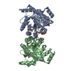 2r6oS C: citing same article ( S: Starting model for refinement |
|---|---|
| Similar structure data |


-Links
 PDBj
PDBj


- Assembly
| Deposited unit | 
| ||||||||
|---|---|---|---|---|---|---|---|---|---|
| 1 |
| ||||||||
| Unit cell |
|
-Components
| #1: Protein | Mass: 30282.393 Da / Num. of mol.: 2 / Fragment: EAL DOMAIN, RESIDUES 439-691 Source method: isolated from a genetically manipulated source Source: (gene. exp.) PSEUDOMONAS AERUGINOSA (bacteria) / Strain: PA01 / Gene: PA4959 / Production host: ESCHERICHIA COLI (E. coli) / Strain (production host): BL21 / Variant (production host): ROSETTA 2References: UniProt: Q9HUK6, cyclic-guanylate-specific phosphodiesterase#2: Chemical | Phosphate  Mass: 94.971 Da / Num. of mol.: 3 / Source method: obtained synthetically / Formula: PO4 Mass: 94.971 Da / Num. of mol.: 3 / Source method: obtained synthetically / Formula: PO4#3: Chemical | ChemComp-ACT / | Acetate  Mass: 59.044 Da / Num. of mol.: 1 / Source method: obtained synthetically / Formula: C2H3O2 Mass: 59.044 Da / Num. of mol.: 1 / Source method: obtained synthetically / Formula: C2H3O2#4: Water | ChemComp-HOH / | Water Mass: 18.015 Da / Num. of mol.: 115 / Source method: isolated from a natural source / Formula: H2O Mass: 18.015 Da / Num. of mol.: 115 / Source method: isolated from a natural source / Formula: H2O |
|---|
-Experimental details
-Experiment
| Experiment | Method: X-RAY DIFFRACTION / Number of used crystals: 1 |
|---|
- Sample preparation
| Crystal | Density Matthews: 2.53 Å3/Da / Density % sol: 51.49 % / Description: NONE |
|---|---|
| Crystal grow | pH: 7 Details: 0.1 M NA ACETATE PH 4.5, 0.4 M K2HPO4, 1.2 M NAH2PO4 |
-Data collection
| Diffraction | Mean temperature: 100 K |
|---|---|
| Diffraction source | Source: SYNCHROTRON / Site: SOLEIL  / Beamline: PROXIMA 1 / Wavelength: 0.9793 / Beamline: PROXIMA 1 / Wavelength: 0.9793 |
| Detector | Type: ADSC QUANTUM 315r / Detector: CCD / Date: Mar 18, 2009 |
| Radiation | Protocol: SINGLE WAVELENGTH / Monochromatic (M) / Laue (L): M / Scattering type: x-ray |
| Radiation wavelength | Wavelength: 0.9793 Å / Relative weight: 1 |
| Reflection | Resolution: 2.3→46.05 Å / Num. obs: 27564 / % possible obs: 99 % / Observed criterion σ(I): 2 / Redundancy: 3.6 % / Biso Wilson estimate: 52.3 Å2 / Rmerge(I) obs: 0.04 / Net I/σ(I): 22.21 |
| Reflection shell | Resolution: 2.3→2.44 Å / Redundancy: 2.7 % / Rmerge(I) obs: 0.43 / Mean I/σ(I) obs: 3.13 / % possible all: 98.2 |
- Processing
| Software |
| |||||||||||||||||||||||||||||||||||||||||||||||||||||||||||||||||||||||||||||
|---|---|---|---|---|---|---|---|---|---|---|---|---|---|---|---|---|---|---|---|---|---|---|---|---|---|---|---|---|---|---|---|---|---|---|---|---|---|---|---|---|---|---|---|---|---|---|---|---|---|---|---|---|---|---|---|---|---|---|---|---|---|---|---|---|---|---|---|---|---|---|---|---|---|---|---|---|---|---|
| Refinement | Method to determine structure: MOLECULAR REPLACEMENT Starting model: PDB ENTRY 2R6O Resolution: 2.304→40.626 Å / SU ML: 0.4 / σ(F): 2 / Phase error: 29.45 / Stereochemistry target values: ML
| |||||||||||||||||||||||||||||||||||||||||||||||||||||||||||||||||||||||||||||
| Solvent computation | Shrinkage radii: 0.98 Å / VDW probe radii: 1.2 Å / Solvent model: FLAT BULK SOLVENT MODEL / Bsol: 52.902 Å2 / ksol: 0.356 e/Å3 | |||||||||||||||||||||||||||||||||||||||||||||||||||||||||||||||||||||||||||||
| Displacement parameters | Biso mean: 57.09 Å2
| |||||||||||||||||||||||||||||||||||||||||||||||||||||||||||||||||||||||||||||
| Refinement step | Cycle: LAST / Resolution: 2.304→40.626 Å
| |||||||||||||||||||||||||||||||||||||||||||||||||||||||||||||||||||||||||||||
| Refine LS restraints |
| |||||||||||||||||||||||||||||||||||||||||||||||||||||||||||||||||||||||||||||
| LS refinement shell |
|
