 Movie
Movie Controller

[English] 日本語
 Yorodumi
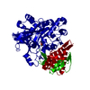
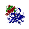
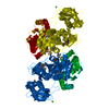
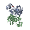
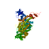
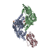
- PDB-2qvx: 4-Chlorobenzoyl-CoA Ligase/Synthetase, I303G mutation, bound to 3... -
+ Open data

- Basic information
| Entry | Database: PDB / ID: 2qvx | ||||||
|---|---|---|---|---|---|---|---|
| Title | 4-Chlorobenzoyl-CoA Ligase/Synthetase, I303G mutation, bound to 3-Chlorobenzoate | ||||||
 Components Components | 4-Chlorobenzoate CoA Ligase | ||||||
 Keywords Keywords |  LIGASE / Adenylate-forming enzymes / Acyl-CoA ligase LIGASE / Adenylate-forming enzymes / Acyl-CoA ligase | ||||||
| Function / homology |  Function and homology information Function and homology informationmedium-chain fatty acid-CoA ligase activity / fatty acid metabolic process / nucleotide binding / membrane / metal ion bindingSimilarity search - Function | ||||||
| Biological species |  Alcaligenes sp. (bacteria) Alcaligenes sp. (bacteria) | ||||||
| Method | X-RAY DIFFRACTION / SYNCHROTRON / 1T5D / Resolution: 2.7 Å | ||||||
 Authors Authors | Wu, R. / Reger, A.S. / Cao, J. / Gulick, A.M. / Dunaway-Mariano, D. | ||||||
 Citation Citation | Journal: Biochemistry / Year: 2007 Title: Rational redesign of the 4-chlorobenzoate binding site of 4-chlorobenzoate: coenzyme a ligase for expanded substrate range. Authors: Wu, R. / Reger, A.S. / Cao, J. / Gulick, A.M. / Dunaway-Mariano, D. | ||||||
| History |
| ||||||
| Remark 999 | SEQUENCE THE AUTHOR MENTIONED THAT THE SEQUENCE USED IN THIS ENTRY MATCHES THE GB ACCESSION CODE ...SEQUENCE THE AUTHOR MENTIONED THAT THE SEQUENCE USED IN THIS ENTRY MATCHES THE GB ACCESSION CODE AAN10109 AND IS MUTATED AT RESIDUE 303 (I303G). |
- Structure visualization
| Structure viewer | Molecule: MolmilJmol/JSmol |
|---|
- Downloads & links
-Download
| PDBx/mmCIF format | 2qvx.cif.gz | 105.7 KB | Display | PDBx/mmCIF format |
|---|---|---|---|---|
| PDB format | pdb2qvx.ent.gz | 81 KB | Display | PDB format |
| PDBx/mmJSON format | 2qvx.json.gz | Tree view | PDBx/mmJSON format | |
| Others |  Other downloads Other downloads |
-Validation report
| Arichive directory | https://data.pdbj.org/pub/pdb/validation_reports/qv/2qvxftp://data.pdbj.org/pub/pdb/validation_reports/qv/2qvx | HTTPS FTP |
|---|
-Related structure data


-Links
 PDBj
PDBj
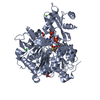
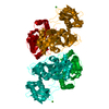
- Assembly
| Deposited unit | 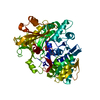
| ||||||||
|---|---|---|---|---|---|---|---|---|---|
| 1 | 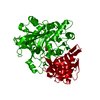
| ||||||||
| Unit cell |
|
-Components
| #1: Protein | Mass: 54325.004 Da / Num. of mol.: 1 Source method: isolated from a genetically manipulated source Source: (gene. exp.) Alcaligenes sp. (bacteria) / Strain: AL3007 / Plasmid: pQE-70 / Production host: Escherichia coli (E. coli) / Strain (production host): JM109 / References: UniProt: Q8GN86*PLUS, EC: 6.2.1.33 |
|---|---|
| #2: Chemical | ChemComp-3BZ / 3-Chlorobenzoic acid  Mass: 156.566 Da / Num. of mol.: 1 / Source method: obtained synthetically / Formula: C7H5ClO2 Mass: 156.566 Da / Num. of mol.: 1 / Source method: obtained synthetically / Formula: C7H5ClO2 |
| #3: Water | ChemComp-HOH / Water Mass: 18.015 Da / Num. of mol.: 31 / Source method: isolated from a natural source / Formula: H2O Mass: 18.015 Da / Num. of mol.: 31 / Source method: isolated from a natural source / Formula: H2O |
-Experimental details
-Experiment
| Experiment | Method: X-RAY DIFFRACTION / Number of used crystals: 1 |
|---|
- Sample preparation
| Crystal | Density Matthews: 3.11 Å3/Da / Density % sol: 60.46 % |
|---|---|
| Crystal grow | Temperature: 277 K / Method: vapor diffusion, hanging drop / pH: 6.7 Details: 14-22% pentaerythritol propoxylate 426, 50 mM BTP, 1 mM ATP, 1 mM 3-CB, pH 6.5-6.75, VAPOR DIFFUSION, HANGING DROP, temperature 277K |
-Data collection
| Diffraction | Mean temperature: 113 K |
|---|---|
| Diffraction source | Source: SYNCHROTRON / Site: CHESS  / Beamline: F2 / Wavelength: 0.9793 Å / Beamline: F2 / Wavelength: 0.9793 Å |
| Detector | Type: ADSC QUANTUM 210 / Detector: CCD / Date: Jul 31, 2005 |
| Radiation | Protocol: SINGLE WAVELENGTH / Monochromatic (M) / Laue (L): M / Scattering type: x-ray |
| Radiation wavelength | Wavelength: 0.9793 Å / Relative weight: 1 |
| Reflection | Resolution: 2.7→30 Å / Num. obs: 18648 / % possible obs: 96.9 % / Redundancy: 6.2 % / Rmerge(I) obs: 0.048 / Net I/σ(I): 15 |
| Reflection shell | Resolution: 2.7→2.79 Å / Rmerge(I) obs: 0.4 / Mean I/σ(I) obs: 2.1 / % possible all: 98.9 |
- Processing
| Software |
| ||||||||||||||||||||||||||||||||||||||||||||||||||||||||||||||||||||||||||||||||||||||||||
|---|---|---|---|---|---|---|---|---|---|---|---|---|---|---|---|---|---|---|---|---|---|---|---|---|---|---|---|---|---|---|---|---|---|---|---|---|---|---|---|---|---|---|---|---|---|---|---|---|---|---|---|---|---|---|---|---|---|---|---|---|---|---|---|---|---|---|---|---|---|---|---|---|---|---|---|---|---|---|---|---|---|---|---|---|---|---|---|---|---|---|---|
| Refinement | Method to determine structure: 1T5D / Resolution: 2.7→30 Å / Cor.coef. Fo:Fc: 0.957 / Cor.coef. Fo:Fc free: 0.9 / SU B: 26.103 / SU ML: 0.256 / TLS residual ADP flag: LIKELY RESIDUAL / Cross valid method: THROUGHOUT / ESU R: 0.638 / ESU R Free: 0.347 / Stereochemistry target values: MAXIMUM LIKELIHOOD / Details: HYDROGENS HAVE BEEN ADDED IN THE RIDING POSITIONS
| ||||||||||||||||||||||||||||||||||||||||||||||||||||||||||||||||||||||||||||||||||||||||||
| Solvent computation | Ion probe radii: 0.8 Å / Shrinkage radii: 0.8 Å / VDW probe radii: 1.2 Å / Solvent model: BABINET MODEL WITH MASK | ||||||||||||||||||||||||||||||||||||||||||||||||||||||||||||||||||||||||||||||||||||||||||
| Displacement parameters | Biso mean: 46.663 Å2
| ||||||||||||||||||||||||||||||||||||||||||||||||||||||||||||||||||||||||||||||||||||||||||
| Refinement step | Cycle: LAST / Resolution: 2.7→30 Å
| ||||||||||||||||||||||||||||||||||||||||||||||||||||||||||||||||||||||||||||||||||||||||||
| Refine LS restraints |
| ||||||||||||||||||||||||||||||||||||||||||||||||||||||||||||||||||||||||||||||||||||||||||
| LS refinement shell | Resolution: 2.7→2.77 Å / Total num. of bins used: 20
| ||||||||||||||||||||||||||||||||||||||||||||||||||||||||||||||||||||||||||||||||||||||||||
| Refinement TLS params. | Method: refined / Refine-ID: X-RAY DIFFRACTION
| ||||||||||||||||||||||||||||||||||||||||||||||||||||||||||||||||||||||||||||||||||||||||||
| Refinement TLS group |
|
