 Movie
Movie Controller

[English] 日本語
 Yorodumi
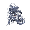
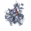
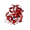
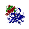
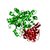
- PDB-1t5h: 4-Chlorobenzoyl-CoA Ligase/Synthetase unliganded, selenomethionine -
+ Open data

- Basic information
| Entry | Database: PDB / ID: 1t5h | ||||||
|---|---|---|---|---|---|---|---|
| Title | 4-Chlorobenzoyl-CoA Ligase/Synthetase unliganded, selenomethionine | ||||||
 Components Components | 4-chlorobenzoyl CoA ligase | ||||||
 Keywords Keywords |  LIGASE / Adenylate-forming coenzyme A Ligase domain alternation conformational change LIGASE / Adenylate-forming coenzyme A Ligase domain alternation conformational change | ||||||
| Function / homology |  Function and homology information Function and homology informationmedium-chain fatty acid-CoA ligase activity / fatty acid metabolic process / nucleotide binding / membrane / metal ion bindingSimilarity search - Function | ||||||
| Biological species |  Alcaligenes sp. AL3007 (bacteria) Alcaligenes sp. AL3007 (bacteria) | ||||||
| Method | X-RAY DIFFRACTION / SYNCHROTRON / MAD / Resolution: 2.002 Å | ||||||
 Authors Authors | Gulick, A.M. / Lu, X. / Dunaway-Mariano, D. | ||||||
 Citation Citation | Journal: Biochemistry / Year: 2004 Title: Crystal Structure of 4-Chlorobenzoate:CoA Ligase/Synthetase in the Unliganded and Aryl Substrate-Bound States Authors: Gulick, A.M. / Lu, X. / Dunaway-Mariano, D. | ||||||
| History |
|
- Structure visualization
| Structure viewer | Molecule: MolmilJmol/JSmol |
|---|
- Downloads & links
-Download
| PDBx/mmCIF format | 1t5h.cif.gz | 110.9 KB | Display | PDBx/mmCIF format |
|---|---|---|---|---|
| PDB format | pdb1t5h.ent.gz | 89.1 KB | Display | PDB format |
| PDBx/mmJSON format | 1t5h.json.gz | Tree view | PDBx/mmJSON format | |
| Others |  Other downloads Other downloads |
-Validation report
| Arichive directory | https://data.pdbj.org/pub/pdb/validation_reports/t5/1t5hftp://data.pdbj.org/pub/pdb/validation_reports/t5/1t5h | HTTPS FTP |
|---|
-Related structure data


-Links
 PDBj
PDBj
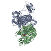
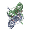
- Assembly
| Deposited unit | 
| ||||||||||
|---|---|---|---|---|---|---|---|---|---|---|---|
| 1 | 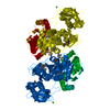
| ||||||||||
| Unit cell |
| ||||||||||
| Components on special symmetry positions |
|
-Components
| #1: Protein | Mass: 54850.059 Da / Num. of mol.: 1 Mutation: SeMet1, SeMet7, SeMet102, SeMet185, SeMet203, SeMet284, SeMet310, SeMet315, SeMet324, SeMet404 Source method: isolated from a genetically manipulated source Source: (gene. exp.) Alcaligenes sp. AL3007 (bacteria) / Plasmid: pQE70 / Production host: Escherichia coli (E. coli) / Strain (production host): JM109 / References: UniProt: Q8GN86, EC: 6.2.1.33 |
|---|---|
| #2: Chemical | ChemComp-CA /   Mass: 40.078 Da / Num. of mol.: 1 / Source method: obtained synthetically / Formula: Ca Mass: 40.078 Da / Num. of mol.: 1 / Source method: obtained synthetically / Formula: Ca |
| #3: Water | ChemComp-HOH / Water Mass: 18.015 Da / Num. of mol.: 340 / Source method: isolated from a natural source / Formula: H2O Mass: 18.015 Da / Num. of mol.: 340 / Source method: isolated from a natural source / Formula: H2O |
-Experimental details
-Experiment
| Experiment | Method: X-RAY DIFFRACTION / Number of used crystals: 1 |
|---|
- Sample preparation
| Crystal | Density Matthews: 2.84 Å3/Da / Density % sol: 56.3 % |
|---|---|
| Crystal grow | Temperature: 277 K / Method: vapor diffusion, hanging drop / pH: 9 Details: 6-8% PEG 4000; 10% glycerol; 0.25 M CaCl2; 50 mM CHES, pH 9.0, VAPOR DIFFUSION, HANGING DROP, temperature 277K |
-Data collection
| Diffraction | Mean temperature: 113 K |
|---|---|
| Diffraction source | Source: SYNCHROTRON / Site: NSLS  / Beamline: X12C / Wavelength: 0.950038 Å / Beamline: X12C / Wavelength: 0.950038 Å |
| Detector | Type: BRANDEIS - B4 / Detector: CCD / Date: Jul 14, 2003 |
| Radiation | Protocol: SINGLE WAVELENGTH / Monochromatic (M) / Laue (L): M / Scattering type: x-ray |
| Radiation wavelength | Wavelength: 0.950038 Å / Relative weight: 1 |
| Reflection | Resolution: 2→25 Å / Num. all: 41528 / Num. obs: 41528 / % possible obs: 100 % / Observed criterion σ(F): 0 / Redundancy: 5.6 % / Biso Wilson estimate: 25.2 Å2 / Rmerge(I) obs: 0.085 / Net I/σ(I): 9.1 |
| Reflection shell | Resolution: 2→2.07 Å / Redundancy: 5.2 % / Rmerge(I) obs: 0.516 / % possible all: 99.7 |
- Processing
| Software |
| |||||||||||||||||||||||||||||||||||||||||||||||||||||||||||||||||||||||||||
|---|---|---|---|---|---|---|---|---|---|---|---|---|---|---|---|---|---|---|---|---|---|---|---|---|---|---|---|---|---|---|---|---|---|---|---|---|---|---|---|---|---|---|---|---|---|---|---|---|---|---|---|---|---|---|---|---|---|---|---|---|---|---|---|---|---|---|---|---|---|---|---|---|---|---|---|---|
| Refinement | Method to determine structure: MAD / Resolution: 2.002→25 Å / Cor.coef. Fo:Fc: 0.955 / Cor.coef. Fo:Fc free: 0.946 / SU B: 3.794 / SU ML: 0.103 / TLS residual ADP flag: LIKELY RESIDUAL / Cross valid method: THROUGHOUT / σ(F): 0 / ESU R: 0.157 / ESU R Free: 0.135 / Stereochemistry target values: MAXIMUM LIKELIHOOD
| |||||||||||||||||||||||||||||||||||||||||||||||||||||||||||||||||||||||||||
| Solvent computation | Ion probe radii: 0.8 Å / Shrinkage radii: 0.8 Å / VDW probe radii: 1.4 Å / Solvent model: BABINET MODEL WITH MASK | |||||||||||||||||||||||||||||||||||||||||||||||||||||||||||||||||||||||||||
| Displacement parameters | Biso mean: 16.44 Å2
| |||||||||||||||||||||||||||||||||||||||||||||||||||||||||||||||||||||||||||
| Refinement step | Cycle: LAST / Resolution: 2.002→25 Å
| |||||||||||||||||||||||||||||||||||||||||||||||||||||||||||||||||||||||||||
| Refine LS restraints |
| |||||||||||||||||||||||||||||||||||||||||||||||||||||||||||||||||||||||||||
| LS refinement shell | Resolution: 2.002→2.053 Å / Total num. of bins used: 20 /
| |||||||||||||||||||||||||||||||||||||||||||||||||||||||||||||||||||||||||||
| Refinement TLS params. | Method: refined / Refine-ID: X-RAY DIFFRACTION
| |||||||||||||||||||||||||||||||||||||||||||||||||||||||||||||||||||||||||||
| Refinement TLS group |
|
