 Movie
Movie Controller


+ Open data

- Basic information
| Entry | Database: PDB / ID: 4aaa | ||||||
|---|---|---|---|---|---|---|---|
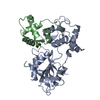
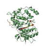
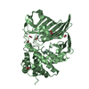
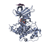
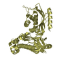
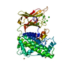
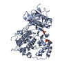
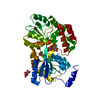
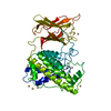
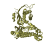
| Title | Crystal structure of the human CDKL2 kinase domain | ||||||
 Components Components | CYCLIN-DEPENDENT KINASE-LIKE 2 | ||||||
 Keywords Keywords |  TRANSFERASE / PHOSPHO-MIMETIC TRANSFERASE / PHOSPHO-MIMETIC | ||||||
| Function / homology |  Function and homology informationsex differentiation / cyclin-dependent kinase / cyclin-dependent protein serine/threonine kinase activity / protein kinase activity / phosphorylation / protein serine kinase activity / protein serine/threonine kinase activity / centrosome / signal transduction / nucleoplasm ...sex differentiation / cyclin-dependent kinase / cyclin-dependent protein serine/threonine kinase activity / protein kinase activity / phosphorylation / protein serine kinase activity / protein serine/threonine kinase activity / centrosome / signal transduction / nucleoplasm / ATP binding / nucleus / cytoplasm Function and homology informationsex differentiation / cyclin-dependent kinase / cyclin-dependent protein serine/threonine kinase activity / protein kinase activity / phosphorylation / protein serine kinase activity / protein serine/threonine kinase activity / centrosome / signal transduction / nucleoplasm ...sex differentiation / cyclin-dependent kinase / cyclin-dependent protein serine/threonine kinase activity / protein kinase activity / phosphorylation / protein serine kinase activity / protein serine/threonine kinase activity / centrosome / signal transduction / nucleoplasm / ATP binding / nucleus / cytoplasmSimilarity search - Function | ||||||
| Biological species |  HOMO SAPIENS (human) HOMO SAPIENS (human) | ||||||
| Method | X-RAY DIFFRACTION / SYNCHROTRON / MOLECULAR REPLACEMENT / Resolution: 1.53 Å | ||||||
 Authors Authors | Canning, P. / Vollmar, M. / Cooper, C.D.O. / Mahajan, P. / Daga, N. / Berridge, G. / Burgess-Brown, N. / Muniz, J.R.C. / Krojer, T. / von Delft, F. ...Canning, P. / Vollmar, M. / Cooper, C.D.O. / Mahajan, P. / Daga, N. / Berridge, G. / Burgess-Brown, N. / Muniz, J.R.C. / Krojer, T. / von Delft, F. / Weigelt, J. / Arrowsmith, C.H. / Edwards, A.M. / Bountra, C. / Bullock, A. | ||||||
 Citation Citation | Journal: Cell Rep / Year: 2018 Title: CDKL Family Kinases Have Evolved Distinct Structural Features and Ciliary Function. Authors: Canning, P. / Park, K. / Goncalves, J. / Li, C. / Howard, C.J. / Sharpe, T.D. / Holt, L.J. / Pelletier, L. / Bullock, A.N. / Leroux, M.R. | ||||||
| History |
|
- Structure visualization
| Structure viewer | Molecule: MolmilJmol/JSmol |
|---|
- Downloads & links
-Download
| PDBx/mmCIF format | 4aaa.cif.gz | 145.4 KB | Display | PDBx/mmCIF format |
|---|---|---|---|---|
| PDB format | pdb4aaa.ent.gz | 114 KB | Display | PDB format |
| PDBx/mmJSON format | 4aaa.json.gz | Tree view | PDBx/mmJSON format | |
| Others |  Other downloads Other downloads |
-Validation report
| Arichive directory | https://data.pdbj.org/pub/pdb/validation_reports/aa/4aaaftp://data.pdbj.org/pub/pdb/validation_reports/aa/4aaa | HTTPS FTP |
|---|
-Related structure data
| Related structure data |  3zduC 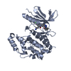 4aguC  4bbmC  4bgqC  3nizS S: Starting model for refinement C: citing same article ( |
|---|---|
| Similar structure data |
-Links
 PDBj
PDBj
- Assembly
| Deposited unit | 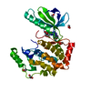
| ||||||||
|---|---|---|---|---|---|---|---|---|---|
| 1 |
| ||||||||
| Unit cell |
|
-Components
| #1: Protein | Mass: 38390.457 Da / Num. of mol.: 1 / Fragment: KINASE DOMAIN, RESIDUES 1-308 / Mutation: YES Source method: isolated from a genetically manipulated source Details: HIS85 CONFORMATION B AND ASP84 CONFORMATION A MAY NOT BE OCCUPIED SIMULTANEOUSLY. Source: (gene. exp.) HOMO SAPIENS (human) / Plasmid: PFB-LIC-BSE / Cell line (production host): SF9 / Production host:   SPODOPTERA FRUGIPERDA (fall armyworm) / References: UniProt: Q92772, cyclin-dependent kinase SPODOPTERA FRUGIPERDA (fall armyworm) / References: UniProt: Q92772, cyclin-dependent kinase | ||||||||
|---|---|---|---|---|---|---|---|---|---|
| #2: Chemical | ChemComp-DKI /   Mass: 425.436 Da / Num. of mol.: 1 / Source method: obtained synthetically / Formula: C15H13F2N7O2S2 Mass: 425.436 Da / Num. of mol.: 1 / Source method: obtained synthetically / Formula: C15H13F2N7O2S2 | ||||||||
| #3: Chemical | ChemComp-EDO / Ethylene glycol  Mass: 62.068 Da / Num. of mol.: 6 / Source method: obtained synthetically / Formula: C2H6O2 Mass: 62.068 Da / Num. of mol.: 6 / Source method: obtained synthetically / Formula: C2H6O2#4: Water | ChemComp-HOH / | Water Mass: 18.015 Da / Num. of mol.: 259 / Source method: isolated from a natural source / Formula: H2O Mass: 18.015 Da / Num. of mol.: 259 / Source method: isolated from a natural source / Formula: H2OCompound details | ENGINEERED | Nonpolymer details | 5-AMINO-3-((4-(AMINOSULFONYL)PHENYL)AMINO)-N-(2, 6-DIFLUOROPHENYL)-1H-1,2,4-TRIAZOLE-1- ...5-AMINO-3-((4-(AMINOSULFO | Sequence details | TWO PHOSPHO-MIMETIC ACTIVATING | |
-Experimental details
-Experiment
| Experiment | Method: X-RAY DIFFRACTION / Number of used crystals: 1 |
|---|
- Sample preparation
| Crystal | Density Matthews: 2.6 Å3/Da / Density % sol: 52.7 % / Description: NONE |
|---|---|
| Crystal grow | Details: 0.2M NACL, 0.1M TRIS PH 8.5; 25% PEG 3350. |
-Data collection
| Diffraction | Mean temperature: 100 K |
|---|---|
| Diffraction source | Source: SYNCHROTRON / Site: Diamond  / Beamline: I02 / Wavelength: 0.9795 / Beamline: I02 / Wavelength: 0.9795 |
| Detector | Type: ADSC CCD / Detector: CCD / Date: Oct 21, 2011 |
| Radiation | Protocol: SINGLE WAVELENGTH / Monochromatic (M) / Laue (L): M / Scattering type: x-ray |
| Radiation wavelength | Wavelength: 0.9795 Å / Relative weight: 1 |
| Reflection | Resolution: 1.6→42.14 Å / Num. obs: 53461 / % possible obs: 100 % / Observed criterion σ(I): 2 / Redundancy: 13.7 % / Rmerge(I) obs: 0.14 / Net I/σ(I): 13.4 |
| Reflection shell | Resolution: 1.6→1.69 Å / Redundancy: 7.9 % / Rmerge(I) obs: 1.14 / Mean I/σ(I) obs: 2.1 / % possible all: 100 |
- Processing
| Software |
| ||||||||||||||||||||||||||||||||||||||||||||||||||||||||||||||||||||||||||||||||||||||||||||||||||||||||||||||||||||||||||||||||||||||||||||||||||||||||||||||||||||||||||||||||||||||
|---|---|---|---|---|---|---|---|---|---|---|---|---|---|---|---|---|---|---|---|---|---|---|---|---|---|---|---|---|---|---|---|---|---|---|---|---|---|---|---|---|---|---|---|---|---|---|---|---|---|---|---|---|---|---|---|---|---|---|---|---|---|---|---|---|---|---|---|---|---|---|---|---|---|---|---|---|---|---|---|---|---|---|---|---|---|---|---|---|---|---|---|---|---|---|---|---|---|---|---|---|---|---|---|---|---|---|---|---|---|---|---|---|---|---|---|---|---|---|---|---|---|---|---|---|---|---|---|---|---|---|---|---|---|---|---|---|---|---|---|---|---|---|---|---|---|---|---|---|---|---|---|---|---|---|---|---|---|---|---|---|---|---|---|---|---|---|---|---|---|---|---|---|---|---|---|---|---|---|---|---|---|---|---|
| Refinement | Method to determine structure: MOLECULAR REPLACEMENT Starting model: PDB ENTRY 3NIZ Resolution: 1.53→53.78 Å / Cor.coef. Fo:Fc: 0.968 / Cor.coef. Fo:Fc free: 0.959 / SU B: 2.88 / SU ML: 0.052 / Cross valid method: THROUGHOUT / ESU R: 0.07 / ESU R Free: 0.073 / Stereochemistry target values: MAXIMUM LIKELIHOOD Details: HYDROGENS HAVE BEEN ADDED IN THE RIDING POSITIONS.U VALUES WITH TLS ADDED. RESIDUES 149 TO 160 ARE DISORDERED RESIDUE HIS85 CONFORMATION B AND ASP84 CONFORMATION A MAY NOT BE OCCUPIED SIMULTANEOUSLY.
| ||||||||||||||||||||||||||||||||||||||||||||||||||||||||||||||||||||||||||||||||||||||||||||||||||||||||||||||||||||||||||||||||||||||||||||||||||||||||||||||||||||||||||||||||||||||
| Solvent computation | Ion probe radii: 0.8 Å / Shrinkage radii: 0.8 Å / VDW probe radii: 1.2 Å / Solvent model: MASK | ||||||||||||||||||||||||||||||||||||||||||||||||||||||||||||||||||||||||||||||||||||||||||||||||||||||||||||||||||||||||||||||||||||||||||||||||||||||||||||||||||||||||||||||||||||||
| Displacement parameters | Biso mean: 24.666 Å2
| ||||||||||||||||||||||||||||||||||||||||||||||||||||||||||||||||||||||||||||||||||||||||||||||||||||||||||||||||||||||||||||||||||||||||||||||||||||||||||||||||||||||||||||||||||||||
| Refinement step | Cycle: LAST / Resolution: 1.53→53.78 Å
| ||||||||||||||||||||||||||||||||||||||||||||||||||||||||||||||||||||||||||||||||||||||||||||||||||||||||||||||||||||||||||||||||||||||||||||||||||||||||||||||||||||||||||||||||||||||
| Refine LS restraints |
|