 Movie
Movie Controller

[English] 日本語
 Yorodumi
- PDB-4a1t: Co-Complex of the of NS3-4A protease with the inhibitory peptide ... -
+ Open data

- Basic information
| Entry | Database: PDB / ID: 4a1t | ||||||
|---|---|---|---|---|---|---|---|
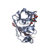
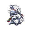
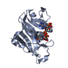
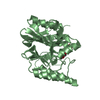
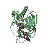
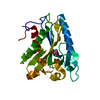
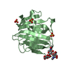
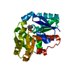
| Title | Co-Complex of the of NS3-4A protease with the inhibitory peptide CP5- 46-A (in-House data) | ||||||
 Components Components |
| ||||||
 Keywords Keywords | HYDROLASE/PEPTIDE / HYDROLASE-PEPTIDE COMPLEX / UNMODIFIED INHIBITORY PEPTIDES | ||||||
| Function / homology |  Function and homology information Function and homology informationRNA stabilization / RNA folding chaperone / DNA/DNA annealing activity / RNA strand annealing activity /  hepacivirin / host cell mitochondrial membrane / host cell lipid droplet / symbiont-mediated suppression of host TRAF-mediated signal transduction / transformation of host cell by virus / symbiont-mediated perturbation of host cell cycle G1/S transition checkpoint ...RNA stabilization / RNA folding chaperone / DNA/DNA annealing activity / RNA strand annealing activity / hepacivirin / host cell mitochondrial membrane / host cell lipid droplet / symbiont-mediated suppression of host TRAF-mediated signal transduction / transformation of host cell by virus / symbiont-mediated perturbation of host cell cycle G1/S transition checkpoint / symbiont-mediated suppression of host JAK-STAT cascade via inhibition of STAT1 activity / symbiont-mediated suppression of host cytoplasmic pattern recognition receptor signaling pathway via inhibition of MAVS activity / protein-DNA complex / : / nucleoside-triphosphate phosphatase / protein complex oligomerization / monoatomic ion channel activity / viral nucleocapsid / clathrin-dependent endocytosis of virus by host cell / Hydrolases; Acting on peptide bonds (peptidases); Cysteine endopeptidases / RNA helicase activity / host cell endoplasmic reticulum membrane / host cell perinuclear region of cytoplasm / RNA helicase / induction by virus of host autophagy / ribonucleoprotein complex / RNA-directed RNA polymerase / viral RNA genome replication / cysteine-type endopeptidase activity / RNA-dependent RNA polymerase activity / serine-type endopeptidase activity / fusion of virus membrane with host endosome membrane / viral envelope / symbiont-mediated suppression of host type I interferon-mediated signaling pathway / host cell nucleus / virion attachment to host cell / host cell plasma membrane / virion membrane / structural molecule activity / ATP hydrolysis activity / proteolysis / DNA binding / RNA binding / zinc ion binding / ATP binding / membrane hepacivirin / host cell mitochondrial membrane / host cell lipid droplet / symbiont-mediated suppression of host TRAF-mediated signal transduction / transformation of host cell by virus / symbiont-mediated perturbation of host cell cycle G1/S transition checkpoint ...RNA stabilization / RNA folding chaperone / DNA/DNA annealing activity / RNA strand annealing activity / hepacivirin / host cell mitochondrial membrane / host cell lipid droplet / symbiont-mediated suppression of host TRAF-mediated signal transduction / transformation of host cell by virus / symbiont-mediated perturbation of host cell cycle G1/S transition checkpoint / symbiont-mediated suppression of host JAK-STAT cascade via inhibition of STAT1 activity / symbiont-mediated suppression of host cytoplasmic pattern recognition receptor signaling pathway via inhibition of MAVS activity / protein-DNA complex / : / nucleoside-triphosphate phosphatase / protein complex oligomerization / monoatomic ion channel activity / viral nucleocapsid / clathrin-dependent endocytosis of virus by host cell / Hydrolases; Acting on peptide bonds (peptidases); Cysteine endopeptidases / RNA helicase activity / host cell endoplasmic reticulum membrane / host cell perinuclear region of cytoplasm / RNA helicase / induction by virus of host autophagy / ribonucleoprotein complex / RNA-directed RNA polymerase / viral RNA genome replication / cysteine-type endopeptidase activity / RNA-dependent RNA polymerase activity / serine-type endopeptidase activity / fusion of virus membrane with host endosome membrane / viral envelope / symbiont-mediated suppression of host type I interferon-mediated signaling pathway / host cell nucleus / virion attachment to host cell / host cell plasma membrane / virion membrane / structural molecule activity / ATP hydrolysis activity / proteolysis / DNA binding / RNA binding / zinc ion binding / ATP binding / membraneSimilarity search - Function | ||||||
| Biological species |  HEPATITIS C VIRUS HEPATITIS C VIRUSSYNTHETIC CONSTRUCT (others) | ||||||
| Method | X-RAY DIFFRACTION / MOLECULAR REPLACEMENT / Resolution: 2.05 Å | ||||||
 Authors Authors | Schmelz, S. / Kuegler, J. / Collins, J. / Heinz, D.W. | ||||||
 Citation Citation | Journal: J.Biol.Chem. / Year: 2012 Title: High Affinity Peptide Inhibitors of the Hepatitis C Virus Ns3-4A Protease Refractory to Common Resistant Mutants. Authors: Kugler, J. / Schmelz, S. / Gentzsch, J. / Haid, S. / Pollmann, E. / Van Den Heuvel, J. / Franke, R. / Pietschmann, T. / Heinz, D.W. / Collins, J. | ||||||
| History |
| ||||||
| Remark 700 | SHEET DETERMINATION METHOD: DSSP THE SHEETS PRESENTED AS "AB" IN EACH CHAIN ON SHEET RECORDS BELOW ... SHEET DETERMINATION METHOD: DSSP THE SHEETS PRESENTED AS "AB" IN EACH CHAIN ON SHEET RECORDS BELOW IS ACTUALLY AN 6-STRANDED BARREL THIS IS REPRESENTED BY A 7-STRANDED SHEET IN WHICH THE FIRST AND LAST STRANDS ARE IDENTICAL. THE SHEETS PRESENTED AS "BB" IN EACH CHAIN ON SHEET RECORDS BELOW IS ACTUALLY AN 6-STRANDED BARREL THIS IS REPRESENTED BY A 7-STRANDED SHEET IN WHICH THE FIRST AND LAST STRANDS ARE IDENTICAL. |
- Structure visualization
| Structure viewer | Molecule: MolmilJmol/JSmol |
|---|
- Downloads & links
-Download
| PDBx/mmCIF format | 4a1t.cif.gz | 99.5 KB | Display | PDBx/mmCIF format |
|---|---|---|---|---|
| PDB format | pdb4a1t.ent.gz | 75.4 KB | Display | PDB format |
| PDBx/mmJSON format | 4a1t.json.gz | Tree view | PDBx/mmJSON format | |
| Others |  Other downloads Other downloads |
-Validation report
| Arichive directory | https://data.pdbj.org/pub/pdb/validation_reports/a1/4a1tftp://data.pdbj.org/pub/pdb/validation_reports/a1/4a1t | HTTPS FTP |
|---|
-Related structure data
| Related structure data | 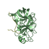 4a1vC 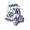 4a1xC 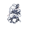 1dxpS S: Starting model for refinement C: citing same article ( |
|---|---|
| Similar structure data |


-Links
 PDBj
PDBj


- Assembly
| Deposited unit | 
| ||||||||
|---|---|---|---|---|---|---|---|---|---|
| 1 | 
| ||||||||
| 2 | 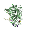
| ||||||||
| Unit cell |
|
-Components
-Protein / Protein/peptide , 2 types, 4 molecules ABCD
| #1: Protein | Mass: 21385.422 Da / Num. of mol.: 2 / Fragment: RESIDUES 1678-1690,1028-1206 Source method: isolated from a genetically manipulated source Details: FUSION PROTEIN OF 4A (1678-1690) WITH NS3 (1028-1206) Source: (gene. exp.) HEPATITIS C VIRUS / Strain: 1B / Description: HCV REPLICON I389/NS3-3'UTR (AJ242654) / Production host:  ESCHERICHIA COLI (E. coli) / Strain (production host): BL21(DE3) / Variant (production host): ROSETTA2 ESCHERICHIA COLI (E. coli) / Strain (production host): BL21(DE3) / Variant (production host): ROSETTA2References: UniProt: P26662, hepacivirin, nucleoside-triphosphate phosphatase, RNA helicase#2: Protein/peptide | Mass: 2304.580 Da / Num. of mol.: 2 / Source method: obtained synthetically / Details: DERIVED FROM PEPTIDE LIBRARY / Source: (synth.) SYNTHETIC CONSTRUCT (others) |
|---|
-Non-polymers , 7 types, 237 molecules 


| #3: Chemical | ChemComp-BCT / Bicarbonate Mass: 61.017 Da / Num. of mol.: 1 / Source method: obtained synthetically / Formula: CHO3 / Comment: pH buffer*YM Mass: 61.017 Da / Num. of mol.: 1 / Source method: obtained synthetically / Formula: CHO3 / Comment: pH buffer*YM | ||||||||||
|---|---|---|---|---|---|---|---|---|---|---|---|
| #4: Chemical | ChemComp-K /  Mass: 39.098 Da / Num. of mol.: 4 / Source method: obtained synthetically / Formula: K Mass: 39.098 Da / Num. of mol.: 4 / Source method: obtained synthetically / Formula: K#5: Chemical | ChemComp-CL / Chloride Mass: 35.453 Da / Num. of mol.: 9 / Source method: obtained synthetically / Formula: Cl Mass: 35.453 Da / Num. of mol.: 9 / Source method: obtained synthetically / Formula: Cl#6: Chemical | Glycerol Mass: 92.094 Da / Num. of mol.: 3 / Source method: obtained synthetically / Formula: C3H8O3 Mass: 92.094 Da / Num. of mol.: 3 / Source method: obtained synthetically / Formula: C3H8O3#7: Chemical |  Mass: 65.409 Da / Num. of mol.: 2 / Source method: obtained synthetically / Formula: Zn Mass: 65.409 Da / Num. of mol.: 2 / Source method: obtained synthetically / Formula: Zn#8: Chemical | ChemComp-DTT / | Dithiothreitol Mass: 154.251 Da / Num. of mol.: 1 / Source method: obtained synthetically / Formula: C4H10O2S2 Mass: 154.251 Da / Num. of mol.: 1 / Source method: obtained synthetically / Formula: C4H10O2S2#9: Water | ChemComp-HOH / | WaterMass: 18.015 Da / Num. of mol.: 217 / Source method: isolated from a natural source / Formula: H2O |
-Details
| Sequence details | SEQUENCE DISCREPANCIES HAVE BEEN INDICATED BY AUTHOR AS NATURAL VARIANTS, AS THE REPLICON WAS ...SEQUENCE DISCREPANC |
|---|
-Experimental details
-Experiment
| Experiment | Method: X-RAY DIFFRACTION / Number of used crystals: 1 |
|---|
- Sample preparation
| Crystal | Density Matthews: 2.32 Å3/Da / Density % sol: 46.91 % / Description: NONE |
|---|---|
| Crystal grow | Details: 0.1 M MES PH 6.0 OR 0.1 M NACITRATE PH 5.1 AND 2.2 M KCL, 5% ISOPROPANOL. |
-Data collection
| Diffraction | Mean temperature: 100 K |
|---|---|
| Diffraction source | Source: ROTATING ANODE / Type: RIGAKU MICROMAX-007 HF / Wavelength: 1.5418 / Wavelength: 1.5418 Å |
| Detector | Type: RIGAKU SATURN 944+ / Detector: CCD / Date: Sep 29, 2010 / Details: MIRRORS |
| Radiation | Protocol: SINGLE WAVELENGTH / Monochromatic (M) / Laue (L): M / Scattering type: x-ray |
| Radiation wavelength | Wavelength: 1.5418 Å / Relative weight: 1 |
| Reflection | Resolution: 2.05→19.7 Å / Num. obs: 25758 / % possible obs: 99.9 % / Observed criterion σ(I): 3.6 / Redundancy: 6.9 % / Biso Wilson estimate: 26.783 Å2 / Rmerge(I) obs: 0.12 / Net I/σ(I): 14.6 |
| Reflection shell | Resolution: 2.05→2.1 Å / Redundancy: 6.7 % / Rmerge(I) obs: 0.6 / Mean I/σ(I) obs: 3.6 / % possible all: 100 |
- Processing
| Software |
| ||||||||||||||||||||||||||||||||||||||||||||||||||||||||||||||||||||||||||||||||||||||||||||||||||||||||||||||||||||||||||||||||||||||||||||||||||||||||||||||||||||||||||||||||||||||
|---|---|---|---|---|---|---|---|---|---|---|---|---|---|---|---|---|---|---|---|---|---|---|---|---|---|---|---|---|---|---|---|---|---|---|---|---|---|---|---|---|---|---|---|---|---|---|---|---|---|---|---|---|---|---|---|---|---|---|---|---|---|---|---|---|---|---|---|---|---|---|---|---|---|---|---|---|---|---|---|---|---|---|---|---|---|---|---|---|---|---|---|---|---|---|---|---|---|---|---|---|---|---|---|---|---|---|---|---|---|---|---|---|---|---|---|---|---|---|---|---|---|---|---|---|---|---|---|---|---|---|---|---|---|---|---|---|---|---|---|---|---|---|---|---|---|---|---|---|---|---|---|---|---|---|---|---|---|---|---|---|---|---|---|---|---|---|---|---|---|---|---|---|---|---|---|---|---|---|---|---|---|---|---|
| Refinement | Method to determine structure: MOLECULAR REPLACEMENT Starting model: PDB ENTRY 1DXP Resolution: 2.05→19.71 Å / Cor.coef. Fo:Fc: 0.929 / Cor.coef. Fo:Fc free: 0.886 / SU B: 4.97 / SU ML: 0.136 / Cross valid method: THROUGHOUT / ESU R: 0.232 / ESU R Free: 0.192 / Stereochemistry target values: MAXIMUM LIKELIHOOD / Details: HYDROGENS HAVE BEEN ADDED IN THE RIDING POSITIONS.
| ||||||||||||||||||||||||||||||||||||||||||||||||||||||||||||||||||||||||||||||||||||||||||||||||||||||||||||||||||||||||||||||||||||||||||||||||||||||||||||||||||||||||||||||||||||||
| Solvent computation | Ion probe radii: 0.8 Å / Shrinkage radii: 0.8 Å / VDW probe radii: 1.2 Å / Solvent model: BABINET MODEL WITH MASK | ||||||||||||||||||||||||||||||||||||||||||||||||||||||||||||||||||||||||||||||||||||||||||||||||||||||||||||||||||||||||||||||||||||||||||||||||||||||||||||||||||||||||||||||||||||||
| Displacement parameters | Biso mean: 23.866 Å2
| ||||||||||||||||||||||||||||||||||||||||||||||||||||||||||||||||||||||||||||||||||||||||||||||||||||||||||||||||||||||||||||||||||||||||||||||||||||||||||||||||||||||||||||||||||||||
| Refinement step | Cycle: LAST / Resolution: 2.05→19.71 Å
| ||||||||||||||||||||||||||||||||||||||||||||||||||||||||||||||||||||||||||||||||||||||||||||||||||||||||||||||||||||||||||||||||||||||||||||||||||||||||||||||||||||||||||||||||||||||
| Refine LS restraints |
|