 Movie
Movie Controller

[English] 日本語
 Yorodumi
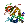
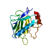
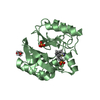
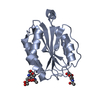
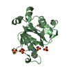
- PDB-3wy8: Crystal Structure of Protease Anisep from Arthrobacter Nicotinovorans -
+ Open data

- Basic information
| Entry | Database: PDB / ID: 3wy8 | ||||||
|---|---|---|---|---|---|---|---|
| Title | Crystal Structure of Protease Anisep from Arthrobacter Nicotinovorans | ||||||
 Components Components | Serine protease | ||||||
 Keywords Keywords | HYDROLASE / TRYPSIN-LIKE | ||||||
| Function / homology | peptidase activity / Trypsin-like serine proteases / Thrombin, subunit H / Peptidase S1, PA clan, chymotrypsin-like fold / Peptidase S1, PA clan / Beta Barrel / Mainly Beta / Serine protease Function and homology information Function and homology information | ||||||
| Biological species |  Arthrobacter nicotinovorans (bacteria) Arthrobacter nicotinovorans (bacteria) | ||||||
| Method | X-RAY DIFFRACTION / SYNCHROTRON / MOLECULAR REPLACEMENT / Resolution: 1.7 Å | ||||||
 Authors Authors | Sone, T. / Haraguchi, Y. / Kuwahara, A. / Ose, T. / Takano, M. / Abe, A. / Tanaka, M. / Tanaka, I. / Asano, K. | ||||||
 Citation Citation | Journal: Protein Pept.Lett. / Year: 2015 Title: Structural characterization reveals the keratinolytic activity of an arthrobacter nicotinovorans protease. Authors: Sone, T. / Haraguchi, Y. / Kuwahara, A. / Ose, T. / Takano, M. / Abe, A. / Tanaka, M. / Tanaka, I. / Asano, K. | ||||||
| History |
|
- Structure visualization
| Structure viewer | Molecule: MolmilJmol/JSmol |
|---|
- Downloads & links
-Download
| PDBx/mmCIF format | 3wy8.cif.gz | 58.8 KB | Display | PDBx/mmCIF format |
|---|---|---|---|---|
| PDB format | pdb3wy8.ent.gz | 42.5 KB | Display | PDB format |
| PDBx/mmJSON format | 3wy8.json.gz | Tree view | PDBx/mmJSON format | |
| Others |  Other downloads Other downloads |
-Validation report
| Arichive directory | https://data.pdbj.org/pub/pdb/validation_reports/wy/3wy8ftp://data.pdbj.org/pub/pdb/validation_reports/wy/3wy8 | HTTPS FTP |
|---|
-Related structure data
| Related structure data |  3cp7S S: Starting model for refinement |
|---|---|
| Similar structure data |


-Links
 PDBj
PDBj- Assembly
| Deposited unit | 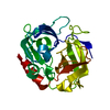
| ||||||||
|---|---|---|---|---|---|---|---|---|---|
| 1 |
| ||||||||
| Unit cell |
| ||||||||
| Components on special symmetry positions |
|
-Components
| #1: Protein | Mass: 22781.861 Da / Num. of mol.: 1 Source method: isolated from a genetically manipulated source Source: (gene. exp.) Arthrobacter nicotinovorans (bacteria) / Strain: 23-0-11 / Gene: Spr / Plasmid: pDEST14 / Production host: Escherichia coli (E. coli) / Strain (production host): BL21-AI / References: UniProt: K7ZK39, chymotrypsin |
|---|---|
| #2: Water | ChemComp-HOH / Water Mass: 18.015 Da / Num. of mol.: 288 / Source method: isolated from a natural source / Formula: H2O Mass: 18.015 Da / Num. of mol.: 288 / Source method: isolated from a natural source / Formula: H2O |
-Experimental details
-Experiment
| Experiment | Method: X-RAY DIFFRACTION / Number of used crystals: 1 |
|---|
- Sample preparation
| Crystal | Density Matthews: 2 Å3/Da / Density % sol: 38.48 % |
|---|---|
| Crystal grow | Temperature: 293 K / Method: vapor diffusion, hanging drop / pH: 7.5 Details: 0.1M HEPES, 4.3M sodium chloride, pH 7.5, VAPOR DIFFUSION, HANGING DROP, temperature 293K |
-Data collection
| Diffraction | Mean temperature: 100 K |
|---|---|
| Diffraction source | Source: SYNCHROTRON / Site: Photon Factory  / Beamline: AR-NW12A / Wavelength: 1 Å / Beamline: AR-NW12A / Wavelength: 1 Å |
| Detector | Type: ADSC QUANTUM 210 / Detector: CCD / Date: Dec 22, 2009 / Details: mirrors |
| Radiation | Monochromator: silicon / Protocol: SINGLE WAVELENGTH / Monochromatic (M) / Laue (L): M / Scattering type: x-ray |
| Radiation wavelength | Wavelength: 1 Å / Relative weight: 1 |
| Reflection | Resolution: 1.7→50 Å / Num. all: 20158 / Num. obs: 20158 / % possible obs: 100 % / Redundancy: 22.2 % / Biso Wilson estimate: 20.68 Å2 / Rmerge(I) obs: 0.076 / Net I/σ(I): 44.9 |
| Reflection shell | Resolution: 1.7→1.73 Å / Redundancy: 22 % / Rmerge(I) obs: 0.5 / Mean I/σ(I) obs: 7.2 / Num. unique all: 1001 / % possible all: 100 |
- Processing
| Software |
| ||||||||||||||||||||||||||||||||||||||||||||||||||||||||||||||||||||||||||||||||||||||||||||||||||||||||||||||||||||||||||||||||||||||||||||||||||||||||||||||||||||||||||||||||||||||
|---|---|---|---|---|---|---|---|---|---|---|---|---|---|---|---|---|---|---|---|---|---|---|---|---|---|---|---|---|---|---|---|---|---|---|---|---|---|---|---|---|---|---|---|---|---|---|---|---|---|---|---|---|---|---|---|---|---|---|---|---|---|---|---|---|---|---|---|---|---|---|---|---|---|---|---|---|---|---|---|---|---|---|---|---|---|---|---|---|---|---|---|---|---|---|---|---|---|---|---|---|---|---|---|---|---|---|---|---|---|---|---|---|---|---|---|---|---|---|---|---|---|---|---|---|---|---|---|---|---|---|---|---|---|---|---|---|---|---|---|---|---|---|---|---|---|---|---|---|---|---|---|---|---|---|---|---|---|---|---|---|---|---|---|---|---|---|---|---|---|---|---|---|---|---|---|---|---|---|---|---|---|---|---|
| Refinement | Method to determine structure: MOLECULAR REPLACEMENT Starting model: 3CP7 Resolution: 1.7→29.74 Å / Cor.coef. Fo:Fc: 0.972 / Cor.coef. Fo:Fc free: 0.962 / SU B: 1.858 / SU ML: 0.062 / Cross valid method: THROUGHOUT / ESU R: 0.108 / ESU R Free: 0.099 / Stereochemistry target values: MAXIMUM LIKELIHOOD
| ||||||||||||||||||||||||||||||||||||||||||||||||||||||||||||||||||||||||||||||||||||||||||||||||||||||||||||||||||||||||||||||||||||||||||||||||||||||||||||||||||||||||||||||||||||||
| Solvent computation | Ion probe radii: 0.8 Å / Shrinkage radii: 0.8 Å / VDW probe radii: 1.4 Å / Solvent model: MASK | ||||||||||||||||||||||||||||||||||||||||||||||||||||||||||||||||||||||||||||||||||||||||||||||||||||||||||||||||||||||||||||||||||||||||||||||||||||||||||||||||||||||||||||||||||||||
| Displacement parameters | Biso mean: 21.719 Å2 | ||||||||||||||||||||||||||||||||||||||||||||||||||||||||||||||||||||||||||||||||||||||||||||||||||||||||||||||||||||||||||||||||||||||||||||||||||||||||||||||||||||||||||||||||||||||
| Refinement step | Cycle: LAST / Resolution: 1.7→29.74 Å
| ||||||||||||||||||||||||||||||||||||||||||||||||||||||||||||||||||||||||||||||||||||||||||||||||||||||||||||||||||||||||||||||||||||||||||||||||||||||||||||||||||||||||||||||||||||||
| Refine LS restraints |
|