 Movie
Movie Controller

[English] 日本語
 Yorodumi
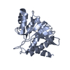
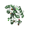
- PDB-6umb: Crystal structure of TRIM7 B30.2 domain at 1.8 angstrom resolution -
+ Open data

- Basic information
| Entry | Database: PDB / ID: 6umb | ||||||
|---|---|---|---|---|---|---|---|
| Title | Crystal structure of TRIM7 B30.2 domain at 1.8 angstrom resolution | ||||||
 Components Components | (E3 ubiquitin-protein ligase TRIM7) x 2 | ||||||
 Keywords Keywords |  LIGASE / B30.2 / E3 ubiquitin ligase / PRY/SPRY / TRIM7 LIGASE / B30.2 / E3 ubiquitin ligase / PRY/SPRY / TRIM7 | ||||||
| Function / homology |  Function and homology information Function and homology informationantiviral innate immune response / RING-type E3 ubiquitin transferase / ubiquitin protein ligase activity / protein ubiquitination / Golgi apparatus / zinc ion binding / nucleus / cytoplasmSimilarity search - Function | ||||||
| Biological species |  Homo sapiens (human) Homo sapiens (human) | ||||||
| Method | X-RAY DIFFRACTION / SYNCHROTRON / MOLECULAR REPLACEMENT / Resolution: 1.8 Å | ||||||
 Authors Authors | Munoz Sosa, C.J. / Carrizo, M.E. | ||||||
 Citation Citation | Journal: J.Biol.Chem. / Year: 2021 Title: Crystal structure and mutational analysis of the human TRIM7 B30.2 domain provide insights into the molecular basis of its binding to glycogenin-1. Authors: Munoz Sosa, C.J. / Issoglio, F.M. / Carrizo, M.E. | ||||||
| History |
|
- Structure visualization
| Structure viewer | Molecule: MolmilJmol/JSmol |
|---|
- Downloads & links
-Download
| PDBx/mmCIF format | 6umb.cif.gz | 88.6 KB | Display | PDBx/mmCIF format |
|---|---|---|---|---|
| PDB format | pdb6umb.ent.gz | 65.5 KB | Display | PDB format |
| PDBx/mmJSON format | 6umb.json.gz | Tree view | PDBx/mmJSON format | |
| Others |  Other downloads Other downloads |
-Validation report
| Arichive directory | https://data.pdbj.org/pub/pdb/validation_reports/um/6umbftp://data.pdbj.org/pub/pdb/validation_reports/um/6umb | HTTPS FTP |
|---|
-Related structure data
| Related structure data | 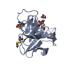 6umaC  4n7iS S: Starting model for refinement C: citing same article ( |
|---|---|
| Similar structure data |


-Links
 PDBj
PDBj


- Assembly
| Deposited unit | 
| ||||||||
|---|---|---|---|---|---|---|---|---|---|
| 1 | 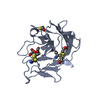
| ||||||||
| 2 | 
| ||||||||
| Unit cell |
|
-Components
| #1: Protein | Mass: 20274.066 Da / Num. of mol.: 1 / Fragment: B30.2 domain Source method: isolated from a genetically manipulated source Source: (gene. exp.) Homo sapiens (human) / Gene: TRIM7, GNIP, RNF90 / Production host:  Escherichia coli (E. coli) Escherichia coli (E. coli)References: UniProt: Q9C029, RING-type E3 ubiquitin transferase | ||||||
|---|---|---|---|---|---|---|---|
| #2: Protein | Mass: 20197.947 Da / Num. of mol.: 1 / Fragment: B30.2 domain Source method: isolated from a genetically manipulated source Source: (gene. exp.) Homo sapiens (human) / Gene: TRIM7, GNIP, RNF90 / Production host: Escherichia coli (E. coli)References: UniProt: Q9C029, RING-type E3 ubiquitin transferase | ||||||
| #3: Chemical | Malonic acid  Mass: 102.046 Da / Num. of mol.: 2 / Source method: obtained synthetically / Formula: C3H2O4 Mass: 102.046 Da / Num. of mol.: 2 / Source method: obtained synthetically / Formula: C3H2O4#4: Chemical | 2-Mercaptoethanol  Mass: 78.133 Da / Num. of mol.: 2 / Source method: obtained synthetically / Formula: C2H6OS Mass: 78.133 Da / Num. of mol.: 2 / Source method: obtained synthetically / Formula: C2H6OS#5: Water | ChemComp-HOH / | Water Mass: 18.015 Da / Num. of mol.: 165 / Source method: isolated from a natural source / Formula: H2O Mass: 18.015 Da / Num. of mol.: 165 / Source method: isolated from a natural source / Formula: H2OHas ligand of interest | N | |
-Experimental details
-Experiment
| Experiment | Method: X-RAY DIFFRACTION / Number of used crystals: 1 |
|---|
- Sample preparation
| Crystal | Density Matthews: 2.29 Å3/Da / Density % sol: 46.25 % |
|---|---|
| Crystal grow | Temperature: 283 K / Method: vapor diffusion, sitting drop / pH: 6 / Details: Sodium malonate pH 6.0 |
-Data collection
| Diffraction | Mean temperature: 100 K / Serial crystal experiment: N | ||||||||||||||||||||||||||||||
|---|---|---|---|---|---|---|---|---|---|---|---|---|---|---|---|---|---|---|---|---|---|---|---|---|---|---|---|---|---|---|---|
| Diffraction source | Source: SYNCHROTRON / Site: LNLS  / Beamline: W01B-MX2 / Wavelength: 1.4586 Å / Beamline: W01B-MX2 / Wavelength: 1.4586 Å | ||||||||||||||||||||||||||||||
| Detector | Type: DECTRIS PILATUS 2M / Detector: PIXEL / Date: Nov 16, 2018 | ||||||||||||||||||||||||||||||
| Radiation | Protocol: SINGLE WAVELENGTH / Monochromatic (M) / Laue (L): M / Scattering type: x-ray | ||||||||||||||||||||||||||||||
| Radiation wavelength | Wavelength: 1.4586 Å / Relative weight: 1 | ||||||||||||||||||||||||||||||
| Reflection | Resolution: 1.8→50.42 Å / Num. obs: 33860 / % possible obs: 99.6 % / Redundancy: 5.7 % / CC1/2: 0.995 / Rmerge(I) obs: 0.076 / Rpim(I) all: 0.035 / Rrim(I) all: 0.084 / Net I/σ(I): 10.7 / Num. measured all: 193627 / Scaling rejects: 1069 | ||||||||||||||||||||||||||||||
| Reflection shell | Diffraction-ID: 1
|
- Processing
| Software |
| ||||||||||||||||||||||||||||||||||||||||||||||||||||||||||||
|---|---|---|---|---|---|---|---|---|---|---|---|---|---|---|---|---|---|---|---|---|---|---|---|---|---|---|---|---|---|---|---|---|---|---|---|---|---|---|---|---|---|---|---|---|---|---|---|---|---|---|---|---|---|---|---|---|---|---|---|---|---|
| Refinement | Method to determine structure: MOLECULAR REPLACEMENT Starting model: 4N7I Resolution: 1.8→45.83 Å / Cor.coef. Fo:Fc: 0.962 / Cor.coef. Fo:Fc free: 0.944 / SU B: 2.918 / SU ML: 0.089 / Cross valid method: THROUGHOUT / σ(F): 0 / ESU R: 0.13 / ESU R Free: 0.125 Details: HYDROGENS HAVE BEEN ADDED IN THE RIDING POSITIONS U VALUES : REFINED INDIVIDUALLY
| ||||||||||||||||||||||||||||||||||||||||||||||||||||||||||||
| Solvent computation | Ion probe radii: 0.8 Å / Shrinkage radii: 0.8 Å / VDW probe radii: 1.2 Å | ||||||||||||||||||||||||||||||||||||||||||||||||||||||||||||
| Displacement parameters | Biso max: 91.85 Å2 / Biso mean: 24.54 Å2 / Biso min: 7.54 Å2
| ||||||||||||||||||||||||||||||||||||||||||||||||||||||||||||
| Refinement step | Cycle: final / Resolution: 1.8→45.83 Å
| ||||||||||||||||||||||||||||||||||||||||||||||||||||||||||||
| Refine LS restraints |
| ||||||||||||||||||||||||||||||||||||||||||||||||||||||||||||
| LS refinement shell | Resolution: 1.804→1.851 Å / Rfactor Rfree error: 0 / Total num. of bins used: 20
|
