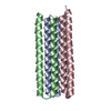
 Movie
Movie Controller


+ Open data

- Basic information
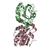
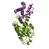
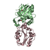
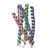
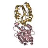
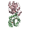
| Entry | Database: PDB / ID: 3wfv | ||||||
|---|---|---|---|---|---|---|---|
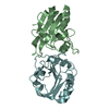
| Title | HIV-1 CRF07 gp41 | ||||||
 Components Components | Envelope glycoprotein gp160 | ||||||
 Keywords Keywords |  VIRAL PROTEIN / DOUBLE HELIX / alpha-helix / virus fusion / glycoprotein / membrane VIRAL PROTEIN / DOUBLE HELIX / alpha-helix / virus fusion / glycoprotein / membrane | ||||||
| Function / homology |  Function and homology information Function and homology informationvirus-mediated perturbation of host defense response => GO:0019049 / : / positive regulation of plasma membrane raft polarization / positive regulation of receptor clustering / positive regulation of establishment of T cell polarity / host cell endosome membrane / clathrin-dependent endocytosis of virus by host cell / membrane => GO:0016020 / viral protein processing / fusion of virus membrane with host plasma membrane ...virus-mediated perturbation of host defense response => GO:0019049 / : / positive regulation of plasma membrane raft polarization / positive regulation of receptor clustering / positive regulation of establishment of T cell polarity / host cell endosome membrane / clathrin-dependent endocytosis of virus by host cell / membrane => GO:0016020 / viral protein processing / fusion of virus membrane with host plasma membrane / fusion of virus membrane with host endosome membrane / viral envelope / virion attachment to host cell / host cell plasma membrane / virion membrane / structural molecule activity / plasma membraneSimilarity search - Function | ||||||
| Biological species |   Human immunodeficiency virus 1 Human immunodeficiency virus 1 | ||||||
| Method | X-RAY DIFFRACTION / SYNCHROTRON / MOLECULAR REPLACEMENT / Resolution: 1.8 Å | ||||||
 Authors Authors | Du, J. / Xue, H. / Ma, J. / Liu, F. / Zhou, J. / Shao, Y. / Qiao, W. / Liu, X. | ||||||
 Citation Citation | Journal: Virology / Year: 2013 Title: The crystal structure of HIV CRF07 B'/C gp41 reveals a hyper-mutant site in the middle of HR2 heptad repeat Authors: Du, J. / Xue, H. / Ma, J. / Liu, F. / Zhou, J. / Shao, Y. / Qiao, W. / Liu, X. | ||||||
| History |
|
- Structure visualization
| Structure viewer | Molecule: MolmilJmol/JSmol |
|---|
- Downloads & links
-Download
| PDBx/mmCIF format | 3wfv.cif.gz | 28.7 KB | Display | PDBx/mmCIF format |
|---|---|---|---|---|
| PDB format | pdb3wfv.ent.gz | 19.1 KB | Display | PDB format |
| PDBx/mmJSON format | 3wfv.json.gz | Tree view | PDBx/mmJSON format | |
| Others |  Other downloads Other downloads |
-Validation report
| Arichive directory | https://data.pdbj.org/pub/pdb/validation_reports/wf/3wfvftp://data.pdbj.org/pub/pdb/validation_reports/wf/3wfv | HTTPS FTP |
|---|
-Related structure data
| Similar structure data |
|---|
-Links
 PDBj
PDBj


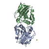
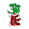
- Assembly
| Deposited unit | 
| ||||||||||||
|---|---|---|---|---|---|---|---|---|---|---|---|---|---|
| 1 | 
| ||||||||||||
| Unit cell |
| ||||||||||||
| Components on special symmetry positions |
|
-Components
| #1: Protein | Mass: 9745.812 Da / Num. of mol.: 1 / Fragment: UNP residues 542-577 and 624-660 Source method: isolated from a genetically manipulated source Source: (gene. exp.) Human immunodeficiency virus 1 / Gene: env / Plasmid: pET30 / Production host:  Escherichia coli (E. coli) / References: UniProt: Q994K2 Escherichia coli (E. coli) / References: UniProt: Q994K2 |
|---|---|
| #2: Water | ChemComp-HOH / Water Mass: 18.015 Da / Num. of mol.: 30 / Source method: isolated from a natural source / Formula: H2O Mass: 18.015 Da / Num. of mol.: 30 / Source method: isolated from a natural source / Formula: H2O |
-Experimental details
-Experiment
| Experiment | Method: X-RAY DIFFRACTION / Number of used crystals: 1 |
|---|
- Sample preparation
| Crystal | Density Matthews: 2.01 Å3/Da / Density % sol: 38.84 % |
|---|---|
| Crystal grow | Temperature: 277 K / Method: vapor diffusion, sitting drop / pH: 8.5 Details: 8% PEG6000, 0.1M Tris, 40% MPD, pH 8.5, VAPOR DIFFUSION, SITTING DROP, temperature 277K |
-Data collection
| Diffraction | Mean temperature: 100 K |
|---|---|
| Diffraction source | Source: SYNCHROTRON / Site: SSRF  / Beamline: BL17U / Wavelength: 0.9793 Å / Beamline: BL17U / Wavelength: 0.9793 Å |
| Detector | Type: ENRAF-NONIUS / Detector: CCD / Date: Sep 16, 2012 |
| Radiation | Monochromator: GRAPHITE / Protocol: SINGLE WAVELENGTH / Monochromatic (M) / Laue (L): M / Scattering type: x-ray |
| Radiation wavelength | Wavelength: 0.9793 Å / Relative weight: 1 |
| Reflection | Resolution: 1.8→20.102 Å / Num. obs: 7326 / % possible obs: 95.76 % / Observed criterion σ(F): 2 / Observed criterion σ(I): 2 |
| Reflection shell | Highest resolution: 1.8 Å / Redundancy: 11.4 % / Rmerge(I) obs: 0.125 / Mean I/σ(I) obs: 17.5 / Num. unique all: 7326 / Rsym value: 0.053 / % possible all: 95.76 |
- Processing
| Software |
| ||||||||||||||||||||||||
|---|---|---|---|---|---|---|---|---|---|---|---|---|---|---|---|---|---|---|---|---|---|---|---|---|---|
| Refinement | Method to determine structure: MOLECULAR REPLACEMENT / Resolution: 1.8→20.102 Å / Occupancy max: 1 / Occupancy min: 1 / FOM work R set: 0.7533 / SU ML: 0.21 / σ(F): 1.38 / Phase error: 29.92 / Stereochemistry target values: ML
| ||||||||||||||||||||||||
| Solvent computation | Shrinkage radii: 0.9 Å / VDW probe radii: 1.11 Å / Solvent model: FLAT BULK SOLVENT MODEL / Bsol: 87.432 Å2 / ksol: 0.478 e/Å3 | ||||||||||||||||||||||||
| Displacement parameters | Biso max: 91.06 Å2 / Biso mean: 32.1715 Å2 / Biso min: 13.76 Å2
| ||||||||||||||||||||||||
| Refinement step | Cycle: LAST / Resolution: 1.8→20.102 Å
| ||||||||||||||||||||||||
| Refine LS restraints |
| ||||||||||||||||||||||||
| LS refinement shell | Refine-ID: X-RAY DIFFRACTION / Total num. of bins used: 2 / % reflection obs: 96 %
|
