 Movie
Movie Controller

[English] 日本語
 Yorodumi
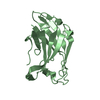
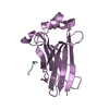
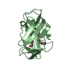
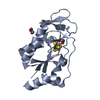
- PDB-3web: Crystal structure of a Niemann-Pick type C2 protein from Japanese... -
+ Open data

- Basic information
| Entry | Database: PDB / ID: 3web | ||||||
|---|---|---|---|---|---|---|---|
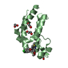
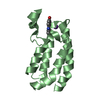
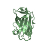
| Title | Crystal structure of a Niemann-Pick type C2 protein from Japanese carpenter ant in complex with oleic acid | ||||||
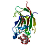
 Components Components | Niemann-Pick type C2 protein | ||||||
 Keywords Keywords | LIPID BINDING PROTEIN / immunoglobulin-like beta-sandwich fold / carrier protein /  Oleic acid Oleic acid | ||||||
| Function / homology |  Function and homology information Function and homology information | ||||||
| Biological species |  Camponotus japonicus (insect) Camponotus japonicus (insect) | ||||||
| Method | X-RAY DIFFRACTION / SYNCHROTRON / MOLECULAR REPLACEMENT / Resolution: 1.7 Å | ||||||
 Authors Authors | Fujimoto, Z. / Tsuchiya, W. / Ishida, Y. / Yamazaki, T. | ||||||
 Citation Citation | Journal: Proc.Natl.Acad.Sci.USA / Year: 2014 Title: Niemann-Pick type C2 protein mediating chemical communication in the worker ant Authors: Ishida, Y. / Tsuchiya, W. / Fujii, T. / Fujimoto, Z. / Miyazawa, M. / Ishibashi, J. / Matsuyama, S. / Ishikawa, Y. / Yamazaki, T. | ||||||
| History |
|
- Structure visualization
| Structure viewer | Molecule: MolmilJmol/JSmol |
|---|
- Downloads & links
-Download
| PDBx/mmCIF format | 3web.cif.gz | 42.4 KB | Display | PDBx/mmCIF format |
|---|---|---|---|---|
| PDB format | pdb3web.ent.gz | 28.3 KB | Display | PDB format |
| PDBx/mmJSON format | 3web.json.gz | Tree view | PDBx/mmJSON format | |
| Others |  Other downloads Other downloads |
-Validation report
| Arichive directory | https://data.pdbj.org/pub/pdb/validation_reports/we/3webftp://data.pdbj.org/pub/pdb/validation_reports/we/3web | HTTPS FTP |
|---|
-Related structure data
| Related structure data | 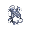 3weaSC S: Starting model for refinement C: citing same article ( |
|---|---|
| Similar structure data |


-Links
 PDBj
PDBj


- Assembly
| Deposited unit | 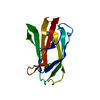
| ||||||||
|---|---|---|---|---|---|---|---|---|---|
| 1 |
| ||||||||
| Unit cell |
|
-Components
| #1: Protein | Mass: 14927.394 Da / Num. of mol.: 1 Source method: isolated from a genetically manipulated source Source: (gene. exp.) Camponotus japonicus (insect) / Gene: CjapNPC2 / Plasmid: pET-22b / Production host:  Escherichia coli (E. coli) / Strain (production host): BL21(DE3) / References: UniProt: W5JXD9*PLUS Escherichia coli (E. coli) / Strain (production host): BL21(DE3) / References: UniProt: W5JXD9*PLUS |
|---|---|
| #2: Chemical | ChemComp-OLA / Oleic acid  Mass: 282.461 Da / Num. of mol.: 1 / Source method: obtained synthetically / Formula: C18H34O2 Mass: 282.461 Da / Num. of mol.: 1 / Source method: obtained synthetically / Formula: C18H34O2 |
| #3: Water | ChemComp-HOH / Water Mass: 18.015 Da / Num. of mol.: 125 / Source method: isolated from a natural source / Formula: H2O Mass: 18.015 Da / Num. of mol.: 125 / Source method: isolated from a natural source / Formula: H2O |
| Sequence details | THE SEQUENCE OF THIS PROTEIN WAS NOT AVAILABLE AT THE UNIPROT KNOWLEDGEBASE DATABASE (UNIPROTKB) AT ...THE SEQUENCE OF THIS PROTEIN WAS NOT AVAILABLE AT THE UNIPROT KNOWLEDGEB |
-Experimental details
-Experiment
| Experiment | Method: X-RAY DIFFRACTION / Number of used crystals: 1 |
|---|
- Sample preparation
| Crystal | Density Matthews: 2.37 Å3/Da / Density % sol: 48.04 % |
|---|---|
| Crystal grow | Temperature: 293 K / Method: vapor diffusion, sitting drop / pH: 7.5 Details: 25% (w/v) PEG 3350, 0.2mM sodium chloride, 0.1M HEPES buffer, pH 7.5, VAPOR DIFFUSION, SITTING DROP, temperature 293K |
-Data collection
| Diffraction | Mean temperature: 95 K |
|---|---|
| Diffraction source | Source: SYNCHROTRON / Site: Photon Factory  / Beamline: BL-5A / Wavelength: 1 Å / Beamline: BL-5A / Wavelength: 1 Å |
| Detector | Type: ADSC QUANTUM 315r / Detector: CCD / Date: Dec 16, 2011 |
| Radiation | Monochromator: Numerical link type Si(111) double crystal monochromator Protocol: SINGLE WAVELENGTH / Monochromatic (M) / Laue (L): M / Scattering type: x-ray |
| Radiation wavelength | Wavelength: 1 Å / Relative weight: 1 |
| Reflection | Resolution: 1.7→31.268 Å / Num. obs: 16234 / % possible obs: 99.9 % / Observed criterion σ(F): 0 / Observed criterion σ(I): -3 / Redundancy: 13.6 % / Biso Wilson estimate: 16.308 Å2 / Rsym value: 0.106 / Net I/σ(I): 16.3 |
| Reflection shell | Resolution: 1.7→1.76 Å / Redundancy: 13.6 % / Mean I/σ(I) obs: 8.2 / Num. unique all: 1576 / Rsym value: 0.443 / % possible all: 100 |
- Processing
| Software |
| |||||||||||||||||||||||||||||||||||||||||||||
|---|---|---|---|---|---|---|---|---|---|---|---|---|---|---|---|---|---|---|---|---|---|---|---|---|---|---|---|---|---|---|---|---|---|---|---|---|---|---|---|---|---|---|---|---|---|---|
| Refinement | Method to determine structure: MOLECULAR REPLACEMENT Starting model: PDB ENTRY 3WEA Resolution: 1.7→31.268 Å / Cor.coef. Fo:Fc: 0.939 / Cor.coef. Fo:Fc free: 0.915 / SU B: 2.48 / SU ML: 0.085 / Cross valid method: THROUGHOUT / ESU R: 0.125 / ESU R Free: 0.128 / Stereochemistry target values: MAXIMUM LIKELIHOOD / Details: HYDROGENS HAVE BEEN USED IF PRESENT IN THE INPUT
| |||||||||||||||||||||||||||||||||||||||||||||
| Solvent computation | Ion probe radii: 0.8 Å / Shrinkage radii: 0.8 Å / VDW probe radii: 1.2 Å / Solvent model: MASK | |||||||||||||||||||||||||||||||||||||||||||||
| Displacement parameters | Biso mean: 22.268 Å2
| |||||||||||||||||||||||||||||||||||||||||||||
| Refine analyze |
| |||||||||||||||||||||||||||||||||||||||||||||
| Refinement step | Cycle: LAST / Resolution: 1.7→31.268 Å
| |||||||||||||||||||||||||||||||||||||||||||||
| Refine LS restraints |
| |||||||||||||||||||||||||||||||||||||||||||||
| LS refinement shell | Resolution: 1.7→1.744 Å / Total num. of bins used: 20
|
