 Movie
Movie Controller


+ Open data

- Basic information
| Entry | Database: PDB / ID: 3w19 | ||||||
|---|---|---|---|---|---|---|---|
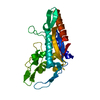
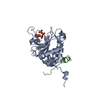
| Title | Potent HIV fusion inhibitor CP32M-2 | ||||||
 Components Components |
| ||||||
 Keywords Keywords |  MEMBRANE PROTEIN/INHIBITOR / 6-helix-bundle / MT-hook / inhibit HIV membrane fusion / MEMBRANE PROTEIN-INHIBITOR complex MEMBRANE PROTEIN/INHIBITOR / 6-helix-bundle / MT-hook / inhibit HIV membrane fusion / MEMBRANE PROTEIN-INHIBITOR complex | ||||||
| Function / homology |  Function and homology information Function and homology informationhost cell periphery / CD4 receptor binding / Dectin-2 family / positive regulation of plasma membrane raft polarization / positive regulation of receptor clustering / positive regulation of establishment of T cell polarity / virus-mediated perturbation of host defense response / host cell endosome membrane / clathrin-dependent endocytosis of virus by host cell / host cell perinuclear region of cytoplasm ...host cell periphery / CD4 receptor binding / Dectin-2 family / positive regulation of plasma membrane raft polarization / positive regulation of receptor clustering / positive regulation of establishment of T cell polarity / virus-mediated perturbation of host defense response / host cell endosome membrane / clathrin-dependent endocytosis of virus by host cell / host cell perinuclear region of cytoplasm / viral protein processing / fusion of virus membrane with host plasma membrane / fusion of virus membrane with host endosome membrane / viral envelope / protein-containing complex binding / virion attachment to host cell / host cell plasma membrane / virion membrane / structural molecule activity / membraneSimilarity search - Function | ||||||
| Biological species |   Human immunodeficiency virus type 1 Human immunodeficiency virus type 1 | ||||||
| Method | X-RAY DIFFRACTION / SYNCHROTRON / MOLECULAR REPLACEMENT / Resolution: 1.278 Å | ||||||
 Authors Authors | Yao, X. / Waltersperger, S. / Wang, M.T. / Cui, S. | ||||||
 Citation Citation | Journal: To be Published Title: Optimization of novel anti-HIV fusion inhibitor Authors: Yao, X. / Waltersperger, S. / Wang, M.T. / Cui, S. | ||||||
| History |
|
- Structure visualization
| Structure viewer | Molecule: MolmilJmol/JSmol |
|---|
- Downloads & links
-Download
| PDBx/mmCIF format | 3w19.cif.gz | 59.1 KB | Display | PDBx/mmCIF format |
|---|---|---|---|---|
| PDB format | pdb3w19.ent.gz | 44.6 KB | Display | PDB format |
| PDBx/mmJSON format | 3w19.json.gz | Tree view | PDBx/mmJSON format | |
| Others |  Other downloads Other downloads |
-Validation report
| Arichive directory | https://data.pdbj.org/pub/pdb/validation_reports/w1/3w19ftp://data.pdbj.org/pub/pdb/validation_reports/w1/3w19 | HTTPS FTP |
|---|
-Related structure data
| Related structure data |  3vgxS S: Starting model for refinement |
|---|---|
| Similar structure data |

-Links
 PDBj
PDBj


- Assembly
| Deposited unit | 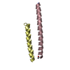
| ||||||||||||
|---|---|---|---|---|---|---|---|---|---|---|---|---|---|
| 1 | 
| ||||||||||||
| Unit cell |
| ||||||||||||
| Components on special symmetry positions |
|
-Components
| #1: Protein/peptide | Transmembrane protein / TM / Glycoprotein 41 / Glycoprotein 120 / gp120 Mass: 4515.291 Da / Num. of mol.: 1 / Fragment: N-peptide T21, UNP RESIDIES 552-589 / Source method: obtained synthetically / Details: THIS SEQUENCE OCCURS NATURALLY IN HIV-1 VIRUS / Source: (synth.) Human immunodeficiency virus type 1 / References: UniProt: P03375 |
|---|---|
| #2: Protein/peptide | Mass: 3825.257 Da / Num. of mol.: 1 / Source method: obtained synthetically Details: THIS SEQUENCE DOES NOT OCCUR NATURALLY IN HIV-1, BUT DESIGNED BASED ON SEQUENCE OF HIV-1 GP41 CHR AND ITS PARENTAL PEPTIDE CP32M |
| #3: Water | ChemComp-HOH / Water Mass: 18.015 Da / Num. of mol.: 75 / Source method: isolated from a natural source / Formula: H2O Mass: 18.015 Da / Num. of mol.: 75 / Source method: isolated from a natural source / Formula: H2O |
-Experimental details
-Experiment
| Experiment | Method: X-RAY DIFFRACTION / Number of used crystals: 1 |
|---|
- Sample preparation
| Crystal | Density Matthews: 2.38 Å3/Da / Density % sol: 48.39 % |
|---|---|
| Crystal grow | Temperature: 295 K / Method: vapor diffusion, hanging drop / pH: 7.5 Details: 0.2M Ammonium sulfate, 0.1 M HEPES, 16%(w/v) PEG 4000, 10%(w/v) isopropanol, pH 7.5, VAPOR DIFFUSION, HANGING DROP, temperature 295K |
-Data collection
| Diffraction | Mean temperature: 100 K |
|---|---|
| Diffraction source | Source: SYNCHROTRON / Site: SLS  / Beamline: X10SA / Wavelength: 0.82656 Å / Beamline: X10SA / Wavelength: 0.82656 Å |
| Detector | Type: PSI PILATUS 6M / Detector: PIXEL / Date: Dec 10, 2011 |
| Radiation | Monochromator: Bartels Monochromator Crystal Type Si (111) / Protocol: SINGLE WAVELENGTH / Monochromatic (M) / Laue (L): M / Scattering type: x-ray |
| Radiation wavelength | Wavelength: 0.82656 Å / Relative weight: 1 |
| Reflection | Resolution: 1.278→36.144 Å / Num. all: 21170 / Num. obs: 21170 / % possible obs: 99.6 % / Observed criterion σ(F): -3 / Observed criterion σ(I): -3 / Redundancy: 5.2 % / Biso Wilson estimate: 13.8 Å2 / Rmerge(I) obs: 0.026 / Net I/σ(I): 27.92 |
| Reflection shell | Resolution: 1.28→1.35 Å / Redundancy: 5.3 % / Rmerge(I) obs: 0.459 / Mean I/σ(I) obs: 3.49 / Num. unique all: 3334 / % possible all: 98.4 |
- Processing
| Software |
| ||||||||||||||||||||||||||||||||||||||||||||||||||||||
|---|---|---|---|---|---|---|---|---|---|---|---|---|---|---|---|---|---|---|---|---|---|---|---|---|---|---|---|---|---|---|---|---|---|---|---|---|---|---|---|---|---|---|---|---|---|---|---|---|---|---|---|---|---|---|---|
| Refinement | Method to determine structure: MOLECULAR REPLACEMENT Starting model: PDB ID 3VGX Resolution: 1.278→36.14 Å / SU ML: 0.15 / Cross valid method: THROUGHOUT / σ(F): 1.46 / Phase error: 14.64 / Stereochemistry target values: ML
| ||||||||||||||||||||||||||||||||||||||||||||||||||||||
| Solvent computation | Shrinkage radii: 0.98 Å / VDW probe radii: 1.2 Å / Solvent model: FLAT BULK SOLVENT MODEL / Bsol: 44.505 Å2 / ksol: 0.347 e/Å3 | ||||||||||||||||||||||||||||||||||||||||||||||||||||||
| Displacement parameters |
| ||||||||||||||||||||||||||||||||||||||||||||||||||||||
| Refinement step | Cycle: LAST / Resolution: 1.278→36.14 Å
| ||||||||||||||||||||||||||||||||||||||||||||||||||||||
| Refine LS restraints |
| ||||||||||||||||||||||||||||||||||||||||||||||||||||||
| LS refinement shell | Refine-ID: X-RAY DIFFRACTION / Total num. of bins used: 8
|
