 Movie
Movie Controller

[English] 日本語
 Yorodumi
- PDB-1we1: Crystal structure of heme oxygenase-1 from cyanobacterium Synecho... -
+ Open data

- Basic information
| Entry | Database: PDB / ID: 1we1 | ||||||
|---|---|---|---|---|---|---|---|
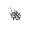
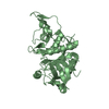
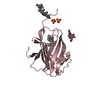
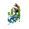
| Title | Crystal structure of heme oxygenase-1 from cyanobacterium Synechocystis sp. PCC6803 in complex with heme | ||||||
 Components Components | Heme oxygenase 1 | ||||||
 Keywords Keywords | OXIDOREDUCTASE | ||||||
| Function / homology |  Function and homology information Function and homology information: / heme oxygenase (biliverdin-producing) / heme oxidation / heme oxygenase (decyclizing) activity / heme catabolic process / photosynthesis / response to oxidative stress / heme binding / metal ion binding Similarity search - Function | ||||||
| Biological species |  | ||||||
| Method |  X-RAY DIFFRACTION / SYNCHROTRON / MOLECULAR REPLACEMENT / Resolution: 2.5 Å X-RAY DIFFRACTION / SYNCHROTRON / MOLECULAR REPLACEMENT / Resolution: 2.5 Å | ||||||
 Authors Authors | Sugishima, M. / Migita, C.T. / Zhang, X. / Yoshida, T. / Fukuyama, K. | ||||||
 Citation Citation | Journal: Eur.J.Biochem. / Year: 2004 Title: Crystal structure of heme oxygenase-1 from cyanobacterium Synechocystis sp. PCC 6803 in complex with heme Authors: Sugishima, M. / Migita, C.T. / Zhang, X. / Yoshida, T. / Fukuyama, K. #1: Journal: Eur.J.Biochem. / Year: 2003 Title: Expression and characterization of cyanobacterium heme oxygenase, a key enzyme in the phycobilin synthesis. Properties of the heme complex of recombinant active enzyme Authors: Migita, C.T. / Zhang, X. / Yoshida, T. | ||||||
| History |
|
- Structure visualization
| Structure viewer | Molecule: MolmilJmol/JSmol |
|---|
- Downloads & links
-Download
| PDBx/mmCIF format | 1we1.cif.gz | 194.6 KB | Display | PDBx/mmCIF format |
|---|---|---|---|---|
| PDB format | pdb1we1.ent.gz | 155.8 KB | Display | PDB format |
| PDBx/mmJSON format | 1we1.json.gz | Tree view | PDBx/mmJSON format | |
| Others |  Other downloads Other downloads |
-Validation report
| Summary document | 1we1_validation.pdf.gz | 1.7 MB | Display | wwPDB validaton report |
|---|---|---|---|---|
| Full document | 1we1_full_validation.pdf.gz | 1.7 MB | Display | |
| Data in XML | 1we1_validation.xml.gz | 40.3 KB | Display | |
| Data in CIF | 1we1_validation.cif.gz | 53.2 KB | Display | |
| Arichive directory | https://data.pdbj.org/pub/pdb/validation_reports/we/1we1ftp://data.pdbj.org/pub/pdb/validation_reports/we/1we1 | HTTPS FTP |
-Related structure data
| Related structure data | 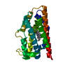 1dveS S: Starting model for refinement |
|---|---|
| Similar structure data |


-Links
 PDBj
PDBj
- Assembly
| Deposited unit | 
| ||||||||
|---|---|---|---|---|---|---|---|---|---|
| 1 | 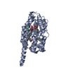
| ||||||||
| 2 | 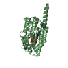
| ||||||||
| 3 | 
| ||||||||
| 4 | 
| ||||||||
| Unit cell |
| ||||||||
| Components on special symmetry positions |
| ||||||||
| Details | biological unit is monomer |
-Components
-Protein , 1 types, 4 molecules ABCD
| #1: Protein | Mass: 27083.664 Da / Num. of mol.: 4 Source method: isolated from a genetically manipulated source Source: (gene. exp.) References: UniProt: P72849, heme oxygenase (biliverdin-producing) |
|---|
-Non-polymers , 5 types, 228 molecules 


| #2: Chemical |  Mass: 94.971 Da / Num. of mol.: 2 / Source method: obtained synthetically / Formula: PO4 Mass: 94.971 Da / Num. of mol.: 2 / Source method: obtained synthetically / Formula: PO4#3: Chemical | ChemComp-CL /  Mass: 35.453 Da / Num. of mol.: 5 / Source method: obtained synthetically / Formula: Cl Mass: 35.453 Da / Num. of mol.: 5 / Source method: obtained synthetically / Formula: Cl#4: Chemical | ChemComp-HEM /  Mass: 616.487 Da / Num. of mol.: 4 / Source method: obtained synthetically / Formula: C34H32FeN4O4 Mass: 616.487 Da / Num. of mol.: 4 / Source method: obtained synthetically / Formula: C34H32FeN4O4#5: Chemical | ChemComp-IPA /  Mass: 60.095 Da / Num. of mol.: 8 / Source method: obtained synthetically / Formula: C3H8O / Comment: alkaloid*YM Mass: 60.095 Da / Num. of mol.: 8 / Source method: obtained synthetically / Formula: C3H8O / Comment: alkaloid*YM#6: Water | ChemComp-HOH / | Mass: 18.015 Da / Num. of mol.: 209 / Source method: isolated from a natural source / Formula: H2O |
|---|
-Experimental details
-Experiment
| Experiment | Method: X-RAY DIFFRACTION / Number of used crystals: 1 |
|---|
- Sample preparation
| Crystal | Density Matthews: 2.9 Å3/Da / Density % sol: 57.1 % |
|---|---|
| Crystal grow | Temperature: 293 K / Method: vapor diffusion, hanging drop / pH: 5.9 Details: PEG 400, 2-propanol, 1,5-diaminopentane dihydrochloride, sodium citrate, pottasium phosphate, pH 5.9, VAPOR DIFFUSION, HANGING DROP, temperature 293K |
-Data collection
| Diffraction | Mean temperature: 100 K |
|---|---|
| Diffraction source | Source: SYNCHROTRON / Site: SPring-8  / Beamline: BL41XU / Wavelength: 1.5 Å / Beamline: BL41XU / Wavelength: 1.5 Å |
| Detector | Type: MARRESEARCH / Detector: CCD / Date: Jan 29, 2004 |
| Radiation | Monochromator: Si(111) double monochromator / Protocol: SINGLE WAVELENGTH / Monochromatic (M) / Laue (L): M / Scattering type: x-ray |
| Radiation wavelength | Wavelength: 1.5 Å / Relative weight: 1 |
| Reflection | Resolution: 2.4→50 Å / Num. all: 46584 / Num. obs: 46584 / % possible obs: 94.7 % / Observed criterion σ(F): 0 / Observed criterion σ(I): 0 / Redundancy: 3.3 % / Biso Wilson estimate: 61.6 Å2 / Rsym value: 0.064 / Net I/σ(I): 7.2 |
| Reflection shell | Resolution: 2.4→2.53 Å / Redundancy: 2.5 % / Mean I/σ(I) obs: 2.4 / Num. unique all: 5929 / Rsym value: 0.302 / % possible all: 83.2 |
- Processing
| Software |
| |||||||||||||||||||||||||
|---|---|---|---|---|---|---|---|---|---|---|---|---|---|---|---|---|---|---|---|---|---|---|---|---|---|---|
| Refinement | Method to determine structure: MOLECULAR REPLACEMENT Starting model: PDB ENTRY 1DVE Resolution: 2.5→20 Å / Isotropic thermal model: isotropic / Cross valid method: THROUGHOUT / σ(F): 0 / Stereochemistry target values: Engh & Huber
| |||||||||||||||||||||||||
| Displacement parameters | Biso mean: -0.079 Å2
| |||||||||||||||||||||||||
| Refine analyze |
| |||||||||||||||||||||||||
| Refinement step | Cycle: LAST / Resolution: 2.5→20 Å
| |||||||||||||||||||||||||
| Refine LS restraints |
| |||||||||||||||||||||||||
| LS refinement shell | Resolution: 2.5→2.59 Å
|
