 Movie
Movie Controller

[English] 日本語
 Yorodumi
- PDB-3vyb: Crystal structure of methyl CpG binding domain of MBD4 in complex... -
+ Open data

- Basic information
| Entry | Database: PDB / ID: 3vyb | ||||||
|---|---|---|---|---|---|---|---|
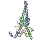
| Title | Crystal structure of methyl CpG binding domain of MBD4 in complex with the 5mCG/hmCG sequence | ||||||
 Components Components |
| ||||||
 Keywords Keywords | HYDROLASE/DNA /  methyl CpG binding domain / protein-DNA complex / versatile base recognition / HYDROLASE-DNA complex methyl CpG binding domain / protein-DNA complex / versatile base recognition / HYDROLASE-DNA complex | ||||||
| Function / homology |  Function and homology information Function and homology informationCleavage of the damaged pyrimidine / Displacement of DNA glycosylase by APEX1 / pyrimidine-specific mismatch base pair DNA N-glycosylase activity / Hydrolases; Glycosylases; Hydrolysing N-glycosyl compounds / mitotic G2 DNA damage checkpoint signaling / response to radiation / intrinsic apoptotic signaling pathway in response to DNA damage / DNA repair / DNA damage response / chromatin ...Cleavage of the damaged pyrimidine / Displacement of DNA glycosylase by APEX1 / pyrimidine-specific mismatch base pair DNA N-glycosylase activity / Hydrolases; Glycosylases; Hydrolysing N-glycosyl compounds / mitotic G2 DNA damage checkpoint signaling / response to radiation / intrinsic apoptotic signaling pathway in response to DNA damage / DNA repair / DNA damage response / chromatin / DNA binding / nucleus / cytoplasmSimilarity search - Function | ||||||
| Biological species |  Mus musculus (house mouse) Mus musculus (house mouse) | ||||||
| Method | X-RAY DIFFRACTION / SYNCHROTRON / MOLECULAR REPLACEMENT / Resolution: 2.4 Å | ||||||
 Authors Authors | Otani, J. / Arita, K. / Kato, T. / Kinoshita, M. / Ariyoshi, M. / Shirakawa, M. | ||||||
 Citation Citation | Journal: J.Biol.Chem. / Year: 2013 Title: Structural basis of the versatile DNA recognition ability of the methyl-CpG binding domain of methyl-CpG binding domain protein 4 Authors: Otani, J. / Arita, K. / Kato, T. / Kinoshita, M. / Kimura, H. / Suetake, I. / Tajima, S. / Ariyoshi, M. / Shirakawa, M. | ||||||
| History |
|
- Structure visualization
| Structure viewer | Molecule: MolmilJmol/JSmol |
|---|
- Downloads & links
-Download
| PDBx/mmCIF format | 3vyb.cif.gz | 75.2 KB | Display | PDBx/mmCIF format |
|---|---|---|---|---|
| PDB format | pdb3vyb.ent.gz | 52.1 KB | Display | PDB format |
| PDBx/mmJSON format | 3vyb.json.gz | Tree view | PDBx/mmJSON format | |
| Others |  Other downloads Other downloads |
-Validation report
| Arichive directory | https://data.pdbj.org/pub/pdb/validation_reports/vy/3vybftp://data.pdbj.org/pub/pdb/validation_reports/vy/3vyb | HTTPS FTP |
|---|
-Related structure data
| Related structure data |  3vxvSC  3vxxC  3vyqC S: Starting model for refinement C: citing same article ( |
|---|---|
| Similar structure data |

-Links
 PDBj
PDBj


- Assembly
| Deposited unit | 
| ||||||||
|---|---|---|---|---|---|---|---|---|---|
| 1 |
| ||||||||
| Unit cell |
| ||||||||
| Components on special symmetry positions |
|
-Components
-Protein , 1 types, 1 molecules A
| #1: Protein | / Methyl-CpG-binding protein MBD4 / Mismatch-specific DNA N-glycosylase Mass: 7829.089 Da / Num. of mol.: 1 / Fragment: methyl CpG binding domain, UNP residues 69-136 Source method: isolated from a genetically manipulated source Source: (gene. exp.) Mus musculus (house mouse) / Gene: Mbd4 / Production host:  Escherichia coli (E. coli) / Strain (production host): BL21(DE3) Escherichia coli (E. coli) / Strain (production host): BL21(DE3)References: UniProt: Q9Z2D7, Hydrolases; Glycosylases; Hydrolysing N-glycosyl compounds |
|---|
-DNA chain , 2 types, 2 molecules BC
| #2: DNA chain | Mass: 4263.809 Da / Num. of mol.: 1 / Source method: obtained synthetically |
|---|---|
| #3: DNA chain | Mass: 4341.827 Da / Num. of mol.: 1 / Source method: obtained synthetically |
-Non-polymers , 3 types, 73 molecules 


| #4: Chemical | ChemComp-ACT / Acetate Mass: 59.044 Da / Num. of mol.: 1 / Source method: obtained synthetically / Formula: C2H3O2 Mass: 59.044 Da / Num. of mol.: 1 / Source method: obtained synthetically / Formula: C2H3O2 |
|---|---|
| #5: Chemical | ChemComp-EDO / Ethylene glycol Mass: 62.068 Da / Num. of mol.: 1 / Source method: obtained synthetically / Formula: C2H6O2 Mass: 62.068 Da / Num. of mol.: 1 / Source method: obtained synthetically / Formula: C2H6O2 |
| #6: Water | ChemComp-HOH / WaterMass: 18.015 Da / Num. of mol.: 71 / Source method: isolated from a natural source / Formula: H2O |
-Experimental details
-Experiment
| Experiment | Method: X-RAY DIFFRACTION / Number of used crystals: 1 |
|---|
- Sample preparation
| Crystal | Density Matthews: 3.67 Å3/Da / Density % sol: 66.53 % |
|---|---|
| Crystal grow | Temperature: 293 K / Method: vapor diffusion, hanging drop / pH: 4 Details: 8% PEG 1500, 0.1M sodium acetate, 0.1M sodium cacodylate, pH 4, VAPOR DIFFUSION, HANGING DROP, temperature 293K |
-Data collection
| Diffraction | Mean temperature: 95 K |
|---|---|
| Diffraction source | Source: SYNCHROTRON / Site: Photon Factory  / Beamline: BL-17A / Wavelength: 1 Å / Beamline: BL-17A / Wavelength: 1 Å |
| Detector | Type: ADSC QUANTUM 315r / Detector: CCD / Date: Oct 8, 2010 |
| Radiation | Protocol: SINGLE WAVELENGTH / Monochromatic (M) / Laue (L): M / Scattering type: x-ray |
| Radiation wavelength | Wavelength: 1 Å / Relative weight: 1 |
| Reflection | Resolution: 2.19→50 Å / Num. obs: 11517 / % possible obs: 89.6 % / Observed criterion σ(F): 1 / Observed criterion σ(I): 1 / Redundancy: 6.5 % / Biso Wilson estimate: 48.25 Å2 / Rmerge(I) obs: 0.052 / Net I/σ(I): 14.7 |
| Reflection shell | Resolution: 2.19→2.27 Å / Redundancy: 4.1 % / Rmerge(I) obs: 0.317 / Num. unique all: 701 / % possible all: 54.7 |
- Processing
| Software |
| ||||||||||||||||||||||||||||||||||||||||||||||||||||||||||||||||||||||||||||||||||||||||||||||||||||
|---|---|---|---|---|---|---|---|---|---|---|---|---|---|---|---|---|---|---|---|---|---|---|---|---|---|---|---|---|---|---|---|---|---|---|---|---|---|---|---|---|---|---|---|---|---|---|---|---|---|---|---|---|---|---|---|---|---|---|---|---|---|---|---|---|---|---|---|---|---|---|---|---|---|---|---|---|---|---|---|---|---|---|---|---|---|---|---|---|---|---|---|---|---|---|---|---|---|---|---|---|---|
| Refinement | Method to determine structure: MOLECULAR REPLACEMENT Starting model: PDB ENTRY 3VXV Resolution: 2.4→34.7 Å / Occupancy max: 1 / Occupancy min: 0.5 / SU ML: 0.43 / σ(F): 0 / Phase error: 24.35 / Stereochemistry target values: ML
| ||||||||||||||||||||||||||||||||||||||||||||||||||||||||||||||||||||||||||||||||||||||||||||||||||||
| Solvent computation | Shrinkage radii: 0.72 Å / VDW probe radii: 1 Å / Solvent model: FLAT BULK SOLVENT MODEL / Bsol: 39.965 Å2 / ksol: 0.329 e/Å3 | ||||||||||||||||||||||||||||||||||||||||||||||||||||||||||||||||||||||||||||||||||||||||||||||||||||
| Displacement parameters | Biso max: 100.75 Å2 / Biso mean: 47.8984 Å2 / Biso min: 22.03 Å2
| ||||||||||||||||||||||||||||||||||||||||||||||||||||||||||||||||||||||||||||||||||||||||||||||||||||
| Refinement step | Cycle: LAST / Resolution: 2.4→34.7 Å
| ||||||||||||||||||||||||||||||||||||||||||||||||||||||||||||||||||||||||||||||||||||||||||||||||||||
| Refine LS restraints |
| ||||||||||||||||||||||||||||||||||||||||||||||||||||||||||||||||||||||||||||||||||||||||||||||||||||
| LS refinement shell | Refine-ID: X-RAY DIFFRACTION / Total num. of bins used: 3
| ||||||||||||||||||||||||||||||||||||||||||||||||||||||||||||||||||||||||||||||||||||||||||||||||||||
| Refinement TLS params. | Method: refined / Refine-ID: X-RAY DIFFRACTION
| ||||||||||||||||||||||||||||||||||||||||||||||||||||||||||||||||||||||||||||||||||||||||||||||||||||
| Refinement TLS group |
|
