 Movie
Movie Controller


+ Open data

- Basic information
| Entry | Database: PDB / ID: 3vtx | ||||||
|---|---|---|---|---|---|---|---|
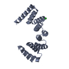
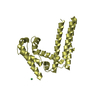
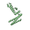
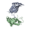
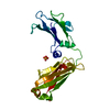
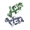
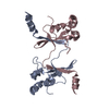
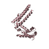
| Title | Crystal structure of MamA protein | ||||||
 Components Components | MamA | ||||||
 Keywords Keywords |  PROTEIN BINDING / Tetratricopeptide repeats (TPR) containing protein / PEPTIDE BINDING PROTEIN PROTEIN BINDING / Tetratricopeptide repeats (TPR) containing protein / PEPTIDE BINDING PROTEIN | ||||||
| Function / homology |  Function and homology informationTetratricopeptide repeat / TPR repeat / Tetratricopeptide repeat 1 / Tetratricopeptide repeat / Tetratricopeptide repeat domain / Tetratricopeptide repeat / TPR repeat region circular profile. / TPR repeat profile. / Tetratricopeptide repeats / Tetratricopeptide repeat ...Tetratricopeptide repeat / TPR repeat / Tetratricopeptide repeat 1 / Tetratricopeptide repeat / Tetratricopeptide repeat domain / Tetratricopeptide repeat / TPR repeat region circular profile. / TPR repeat profile. / Tetratricopeptide repeats / Tetratricopeptide repeat / Serine Threonine Protein Phosphatase 5, Tetratricopeptide repeat / Alpha Horseshoe / Tetratricopeptide-like helical domain superfamily / Mainly Alpha Function and homology informationTetratricopeptide repeat / TPR repeat / Tetratricopeptide repeat 1 / Tetratricopeptide repeat / Tetratricopeptide repeat domain / Tetratricopeptide repeat / TPR repeat region circular profile. / TPR repeat profile. / Tetratricopeptide repeats / Tetratricopeptide repeat ...Tetratricopeptide repeat / TPR repeat / Tetratricopeptide repeat 1 / Tetratricopeptide repeat / Tetratricopeptide repeat domain / Tetratricopeptide repeat / TPR repeat region circular profile. / TPR repeat profile. / Tetratricopeptide repeats / Tetratricopeptide repeat / Serine Threonine Protein Phosphatase 5, Tetratricopeptide repeat / Alpha Horseshoe / Tetratricopeptide-like helical domain superfamily / Mainly AlphaSimilarity search - Domain/homology | ||||||
| Biological species |  Candidatus Magnetobacterium bavaricum (bacteria) Candidatus Magnetobacterium bavaricum (bacteria) | ||||||
| Method | X-RAY DIFFRACTION / SYNCHROTRON / MOLECULAR REPLACEMENT / molecular replacement / Resolution: 1.75 Å | ||||||
 Authors Authors | Zeytuni, N. / Baran, D. / Davidov, G. / Zarivach, R. | ||||||
 Citation Citation | Journal: J.Struct.Biol. / Year: 2012 Title: Inter-phylum structural conservation of the magnetosome-associated TPR-containing protein, MamA Authors: Zeytuni, N. / Baran, D. / Davidov, G. / Zarivach, R. | ||||||
| History |
|
- Structure visualization
| Structure viewer | Molecule: MolmilJmol/JSmol |
|---|
- Downloads & links
-Download
| PDBx/mmCIF format | 3vtx.cif.gz | 168.5 KB | Display | PDBx/mmCIF format |
|---|---|---|---|---|
| PDB format | pdb3vtx.ent.gz | 136.3 KB | Display | PDB format |
| PDBx/mmJSON format | 3vtx.json.gz | Tree view | PDBx/mmJSON format | |
| Others |  Other downloads Other downloads |
-Validation report
| Arichive directory | https://data.pdbj.org/pub/pdb/validation_reports/vt/3vtxftp://data.pdbj.org/pub/pdb/validation_reports/vt/3vtx | HTTPS FTP |
|---|
-Related structure data


-Links
 PDBj
PDBj


- Assembly
| Deposited unit | 
| ||||||||
|---|---|---|---|---|---|---|---|---|---|
| 1 | 
| ||||||||
| 2 | 
| ||||||||
| 3 | 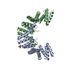
| ||||||||
| Unit cell |
|
-Components
| #1: Protein | Mass: 20648.771 Da / Num. of mol.: 2 Source method: isolated from a genetically manipulated source Source: (gene. exp.) Candidatus Magnetobacterium bavaricum (bacteria)Strain: Mbav / Gene: MamA / Plasmid: pET52b / Production host: Escherichia coli (E. coli) / Strain (production host): BL21(DE3) / References: UniProt: K7N5L8*PLUS#2: Chemical | Glycerol  Mass: 92.094 Da / Num. of mol.: 2 / Source method: obtained synthetically / Formula: C3H8O3 Mass: 92.094 Da / Num. of mol.: 2 / Source method: obtained synthetically / Formula: C3H8O3#3: Water | ChemComp-HOH / | Water Mass: 18.015 Da / Num. of mol.: 376 / Source method: isolated from a natural source / Formula: H2O Mass: 18.015 Da / Num. of mol.: 376 / Source method: isolated from a natural source / Formula: H2OSequence details | THE SEQUENCE DATABASE OF THE THE SEQUENCE OF THIS PROTEIN WAS NOT AVAILABLE AT THE UNIPROT ...THE SEQUENCE DATABASE OF THE THE SEQUENCE OF THIS PROTEIN WAS NOT AVAILABLE AT THE UNIPROT KNOWLEDGEB | |
|---|
-Experimental details
-Experiment
| Experiment | Method: X-RAY DIFFRACTION / Number of used crystals: 1 |
|---|
- Sample preparation
| Crystal | Density Matthews: 2.83 Å3/Da / Density % sol: 56.61 % |
|---|---|
| Crystal grow | Temperature: 293 K / Method: vappor diffusion, sitting drop / pH: 4.4 Details: 0.1M BICINE pH 9, 2M MgCl, Vappor diffusion, sitting drop, temperature 293K |
-Data collection
| Diffraction | Mean temperature: 100 K |
|---|---|
| Diffraction source | Source: SYNCHROTRON / Site: ESRF  / Beamline: ID14-4 / Wavelength: 0.939 Å / Beamline: ID14-4 / Wavelength: 0.939 Å |
| Detector | Type: ADSC QUANTUM 315r / Detector: CCD / Date: Nov 22, 2011 |
| Radiation | Protocol: SINGLE WAVELENGTH / Monochromatic (M) / Laue (L): M / Scattering type: x-ray |
| Radiation wavelength | Wavelength: 0.939 Å / Relative weight: 1 |
| Reflection | Resolution: 1.745→54.98 Å / Num. all: 47365 / Num. obs: 47254 |
| Reflection shell | Resolution: 1.74→1.791 Å |
-Phasing
| Phasing | Method: molecular replacement |
|---|
- Processing
| Software |
| |||||||||||||||||||||||||||||||||||||||||||||||||||||||||||||||||||||||||||||||||||||
|---|---|---|---|---|---|---|---|---|---|---|---|---|---|---|---|---|---|---|---|---|---|---|---|---|---|---|---|---|---|---|---|---|---|---|---|---|---|---|---|---|---|---|---|---|---|---|---|---|---|---|---|---|---|---|---|---|---|---|---|---|---|---|---|---|---|---|---|---|---|---|---|---|---|---|---|---|---|---|---|---|---|---|---|---|---|---|
| Refinement | Method to determine structure: MOLECULAR REPLACEMENT / Resolution: 1.75→54.98 Å / Cor.coef. Fo:Fc: 0.967 / Cor.coef. Fo:Fc free: 0.953 / WRfactor Rfree: 0.2025 / WRfactor Rwork: 0.1618 / Occupancy max: 1 / Occupancy min: 0.5 / FOM work R set: 0.8789 / SU B: 4.057 / SU ML: 0.059 / SU R Cruickshank DPI: 0.094 / SU Rfree: 0.0977 / Cross valid method: THROUGHOUT / σ(F): 0 / ESU R: 0.094 / ESU R Free: 0.098 / Stereochemistry target values: MAXIMUM LIKELIHOOD Details: HYDROGENS HAVE BEEN ADDED IN THE RIDING POSITIONS U VALUES: WITH TLS ADDED
| |||||||||||||||||||||||||||||||||||||||||||||||||||||||||||||||||||||||||||||||||||||
| Solvent computation | Ion probe radii: 0.8 Å / Shrinkage radii: 0.8 Å / VDW probe radii: 1.4 Å / Solvent model: MASK | |||||||||||||||||||||||||||||||||||||||||||||||||||||||||||||||||||||||||||||||||||||
| Displacement parameters | Biso max: 68.77 Å2 / Biso mean: 29.1098 Å2 / Biso min: 12.27 Å2
| |||||||||||||||||||||||||||||||||||||||||||||||||||||||||||||||||||||||||||||||||||||
| Refinement step | Cycle: LAST / Resolution: 1.75→54.98 Å
| |||||||||||||||||||||||||||||||||||||||||||||||||||||||||||||||||||||||||||||||||||||
| Refine LS restraints |
| |||||||||||||||||||||||||||||||||||||||||||||||||||||||||||||||||||||||||||||||||||||
| LS refinement shell | Resolution: 1.746→1.791 Å / Total num. of bins used: 20
| |||||||||||||||||||||||||||||||||||||||||||||||||||||||||||||||||||||||||||||||||||||
| Refinement TLS params. | Method: refined / Refine-ID: X-RAY DIFFRACTION
| |||||||||||||||||||||||||||||||||||||||||||||||||||||||||||||||||||||||||||||||||||||
| Refinement TLS group |
|
