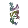
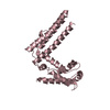
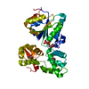
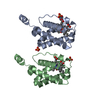
Deposited unit
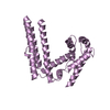
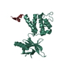
E: ULP_PROTEASE domain-containing protein
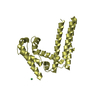
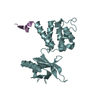
D: ULP_PROTEASE domain-containing protein
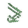
B: ULP_PROTEASE domain-containing protein
F: ULP_PROTEASE domain-containing protein
C: ULP_PROTEASE domain-containing protein
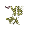
A: ULP_PROTEASE domain-containing protein
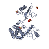
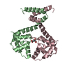
hetero molecules Summary Component details
Theoretical mass Number of molelcules Total (without water) 151,202 7 Polymers 151,143 6 Non-polymers 59 1 Water 18 1
1
E: ULP_PROTEASE domain-containing protein Summary Component details Symmetry operations Calculated values
Theoretical mass Number of molelcules Total (without water) 25,191 1 Polymers 25,191 1 Non-polymers 0 0 Water 0
Type Name Symmetry operation Number identity operation 1_555 x,y,z 1
2
D: ULP_PROTEASE domain-containing protein
hetero molecules Summary Component details Symmetry operations Calculated values
Theoretical mass Number of molelcules Total (without water) 25,249 2 Polymers 25,191 1 Non-polymers 59 1 Water 18 1
Type Name Symmetry operation Number identity operation 1_555 x,y,z 1
3
B: ULP_PROTEASE domain-containing protein Summary Component details Symmetry operations Calculated values
Theoretical mass Number of molelcules Total (without water) 25,191 1 Polymers 25,191 1 Non-polymers 0 0 Water 0
Type Name Symmetry operation Number identity operation 1_555 x,y,z 1
4
F: ULP_PROTEASE domain-containing protein Summary Component details Symmetry operations Calculated values
Theoretical mass Number of molelcules Total (without water) 25,191 1 Polymers 25,191 1 Non-polymers 0 0 Water 0
Type Name Symmetry operation Number identity operation 1_555 x,y,z 1
5
C: ULP_PROTEASE domain-containing protein Summary Component details Symmetry operations Calculated values
Theoretical mass Number of molelcules Total (without water) 25,191 1 Polymers 25,191 1 Non-polymers 0 0 Water 0
Type Name Symmetry operation Number identity operation 1_555 x,y,z 1
6
A: ULP_PROTEASE domain-containing protein Summary Component details Symmetry operations Calculated values
Theoretical mass Number of molelcules Total (without water) 25,191 1 Polymers 25,191 1 Non-polymers 0 0 Water 0
Type Name Symmetry operation Number identity operation 1_555 x,y,z 1
Unit cell Length a, b, c (Å) 110.533, 110.533, 251.240 Angle α, β, γ (deg.) 90.000, 90.000, 120.000 Int Tables number 173 Space group name H-M P63
Components on special symmetry positions ID Model Components 1 1 D -801-NI
Noncrystallographic symmetry (NCS) NCS domain Show large table (3 x 30) Hide large table ID Ens-ID Details 1 1 E2 1 D1 2 E2 2 B1 3 E2 3 F1 4 E2 4 C1 5 E2 5 A1 6 D2 6 B1 7 D2 7 F1 8 D2 8 C1 9 D2 9 A1 10 B2 10 F1 11 B2 11 C1 12 B2 12 A1 13 F2 13 C1 14 F2 14 A1 15 C2 15 A
NCS domain segments Component-ID / Beg auth comp-ID / Beg label comp-ID / Refine code
Show large table (8 x 30) Hide large table Dom-ID Ens-ID End auth comp-ID End label comp-ID Auth asym-ID Label asym-ID Auth seq-ID Label seq-ID 1 1 HISHISEA549 - 762 3 - 216 2 1 HISHISDB549 - 762 3 - 216 1 2 LEULEUEA549 - 758 3 - 212 2 2 LEULEUBC549 - 758 3 - 212 1 3 HISHISEA549 - 760 3 - 214 2 3 HISHISFD549 - 760 3 - 214 1 4 HISHISEA549 - 760 3 - 214 2 4 HISHISCE549 - 760 3 - 214 1 5 LEULEUEA549 - 758 3 - 212 2 5 LEULEUAF549 - 758 3 - 212 1 6 LEULEUDB549 - 758 3 - 212 2 6 LEULEUBC549 - 758 3 - 212 1 7 HISHISDB549 - 760 3 - 214 2 7 HISHISFD549 - 760 3 - 214 1 8 HISHISDB549 - 760 3 - 214 2 8 HISHISCE549 - 760 3 - 214 1 9 LEULEUDB549 - 758 3 - 212 2 9 LEULEUAF549 - 758 3 - 212 1 10 LEULEUBC
 Movie
Movie Controller

 Yorodumi
 Open data
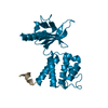
 Basic information
 Components
 Keywords
 SIGNALING PROTEIN /
  Function and homology information
 Authors
 United States, 1items
  Citation
 Structure visualization
 Downloads & links
 Other downloads

 PDBj
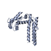
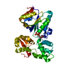
PDBj Assembly