 Movie
Movie Controller


+ Open data

- Basic information
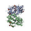
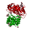
| Entry | Database: PDB / ID: 3itw | ||||||
|---|---|---|---|---|---|---|---|
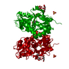
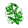
| Title | Crystal structure of TioX from Micromonospora sp. ML1 | ||||||
 Components Components | Protein tioX | ||||||
 Keywords Keywords | PEPTIDE BINDING PROTEIN / bleomycin resistance fold / bisintercalator / solvent-exposed Trp residue / thiocoraline /  PROTEIN BINDING PROTEIN BINDING | ||||||
| Function / homology |  Function and homology information Function and homology informationSignal recognition particle alu RNA binding heterodimer, srp9/1 - #120 / Signal recognition particle alu RNA binding heterodimer, srp9/1 - #110 / Signal recognition particle alu RNA binding heterodimer, srp9/1 / Glyoxalase/fosfomycin resistance/dioxygenase domain / Glyoxalase/Bleomycin resistance protein/Dioxygenase superfamily / Vicinal oxygen chelate (VOC) domain / Vicinal oxygen chelate (VOC) domain profile. / Glyoxalase/Bleomycin resistance protein/Dihydroxybiphenyl dioxygenase / 2-Layer Sandwich / Alpha Beta Similarity search - Domain/homology | ||||||
| Biological species |  Micromonospora sp. ML1 (bacteria) Micromonospora sp. ML1 (bacteria) | ||||||
| Method | X-RAY DIFFRACTION / SYNCHROTRON / Se MAD/MIR / Resolution: 2.15 Å | ||||||
 Authors Authors | Biswas, T. / Garneau-Tsodikova, S. / Tsodikov, O.V. | ||||||
 Citation Citation | Journal: J.Mol.Biol. / Year: 2010 Title: A New Scaffold of an Old Protein Fold Ensures Binding to the Bisintercalator Thiocoraline. Authors: Biswas, T. / Zolova, O.E. / Lombo, F. / de la Calle, F. / Salas, J.A. / Tsodikov, O.V. / Garneau-Tsodikova, S. | ||||||
| History |
|
- Structure visualization
| Structure viewer | Molecule: MolmilJmol/JSmol |
|---|
- Downloads & links
-Download
| PDBx/mmCIF format | 3itw.cif.gz | 58.2 KB | Display | PDBx/mmCIF format |
|---|---|---|---|---|
| PDB format | pdb3itw.ent.gz | 46.9 KB | Display | PDB format |
| PDBx/mmJSON format | 3itw.json.gz | Tree view | PDBx/mmJSON format | |
| Others |  Other downloads Other downloads |
-Validation report
| Arichive directory | https://data.pdbj.org/pub/pdb/validation_reports/it/3itwftp://data.pdbj.org/pub/pdb/validation_reports/it/3itw | HTTPS FTP |
|---|
-Related structure data
| Similar structure data |
|---|

-Links
 PDBj
PDBj- Assembly
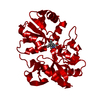
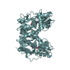
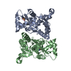
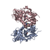
| Deposited unit | 
| ||||||||
|---|---|---|---|---|---|---|---|---|---|
| 1 | 
| ||||||||
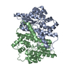
| Unit cell |
|
-Components
| #1: Protein | Mass: 15470.679 Da / Num. of mol.: 2 Source method: isolated from a genetically manipulated source Source: (gene. exp.) Micromonospora sp. ML1 (bacteria) / Gene: tioX / Plasmid: pET28a / Production host: Escherichia coli (E. coli) / Strain (production host): BL21(DE3) / References: UniProt: Q333U2#2: Water | ChemComp-HOH / | Water Mass: 18.015 Da / Num. of mol.: 101 / Source method: isolated from a natural source / Formula: H2O Mass: 18.015 Da / Num. of mol.: 101 / Source method: isolated from a natural source / Formula: H2O |
|---|
-Experimental details
-Experiment
| Experiment | Method: X-RAY DIFFRACTION |
|---|
- Sample preparation
| Crystal | Density Matthews: 3.46 Å3/Da / Density % sol: 64.45 % |
|---|---|
| Crystal grow | Temperature: 295 K / Method: vapor diffusion, hanging drop / pH: 8 Details: Reservoir solution: 400 mM sodium potassium tartrate, 25 mM ammonium sulfate, 2% glycerol, pH 8.0, VAPOR DIFFUSION, HANGING DROP, temperature 295K |
-Data collection
| Diffraction |
| ||||||||||||
|---|---|---|---|---|---|---|---|---|---|---|---|---|---|
| Diffraction source |
| ||||||||||||
| Detector | Date: Aug 16, 2007 | ||||||||||||
| Radiation | Protocol: SINGLE WAVELENGTH / Monochromatic (M) / Laue (L): M / Scattering type: x-ray | ||||||||||||
| Radiation wavelength | Relative weight: 1 | ||||||||||||
| Reflection | Resolution: 2.15→40 Å / Num. obs: 47329 |

- Processing
| Software |
| ||||||||||||||||||||||||||||||||||||||||||||||||||||||||||||||||||||||||||||||||||||||||||||||||||||||||||||||||||||||||||||||||||||||||||||||||||||||||||||||||||||||||||
|---|---|---|---|---|---|---|---|---|---|---|---|---|---|---|---|---|---|---|---|---|---|---|---|---|---|---|---|---|---|---|---|---|---|---|---|---|---|---|---|---|---|---|---|---|---|---|---|---|---|---|---|---|---|---|---|---|---|---|---|---|---|---|---|---|---|---|---|---|---|---|---|---|---|---|---|---|---|---|---|---|---|---|---|---|---|---|---|---|---|---|---|---|---|---|---|---|---|---|---|---|---|---|---|---|---|---|---|---|---|---|---|---|---|---|---|---|---|---|---|---|---|---|---|---|---|---|---|---|---|---|---|---|---|---|---|---|---|---|---|---|---|---|---|---|---|---|---|---|---|---|---|---|---|---|---|---|---|---|---|---|---|---|---|---|---|---|---|---|---|---|---|
| Refinement | Method to determine structure: Se MAD/MIR / Resolution: 2.15→40 Å / Cor.coef. Fo:Fc: 0.944 / Cor.coef. Fo:Fc free: 0.922 / SU B: 11.843 / SU ML: 0.153 / TLS residual ADP flag: LIKELY RESIDUAL / Cross valid method: THROUGHOUT / ESU R: 0.194 / ESU R Free: 0.178 / Stereochemistry target values: MAXIMUM LIKELIHOOD / Details: HYDROGENS HAVE BEEN ADDED IN THE RIDING POSITIONS
| ||||||||||||||||||||||||||||||||||||||||||||||||||||||||||||||||||||||||||||||||||||||||||||||||||||||||||||||||||||||||||||||||||||||||||||||||||||||||||||||||||||||||||
| Solvent computation | Ion probe radii: 0.8 Å / Shrinkage radii: 0.8 Å / VDW probe radii: 1.2 Å / Solvent model: MASK | ||||||||||||||||||||||||||||||||||||||||||||||||||||||||||||||||||||||||||||||||||||||||||||||||||||||||||||||||||||||||||||||||||||||||||||||||||||||||||||||||||||||||||
| Displacement parameters | Biso mean: 73.024 Å2
| ||||||||||||||||||||||||||||||||||||||||||||||||||||||||||||||||||||||||||||||||||||||||||||||||||||||||||||||||||||||||||||||||||||||||||||||||||||||||||||||||||||||||||
| Refinement step | Cycle: LAST / Resolution: 2.15→40 Å
| ||||||||||||||||||||||||||||||||||||||||||||||||||||||||||||||||||||||||||||||||||||||||||||||||||||||||||||||||||||||||||||||||||||||||||||||||||||||||||||||||||||||||||
| Refine LS restraints |
| ||||||||||||||||||||||||||||||||||||||||||||||||||||||||||||||||||||||||||||||||||||||||||||||||||||||||||||||||||||||||||||||||||||||||||||||||||||||||||||||||||||||||||
| LS refinement shell | Resolution: 2.15→2.206 Å / Total num. of bins used: 20
| ||||||||||||||||||||||||||||||||||||||||||||||||||||||||||||||||||||||||||||||||||||||||||||||||||||||||||||||||||||||||||||||||||||||||||||||||||||||||||||||||||||||||||
| Refinement TLS params. | Method: refined / Refine-ID: X-RAY DIFFRACTION
| ||||||||||||||||||||||||||||||||||||||||||||||||||||||||||||||||||||||||||||||||||||||||||||||||||||||||||||||||||||||||||||||||||||||||||||||||||||||||||||||||||||||||||
| Refinement TLS group |
|
