 Movie
Movie Controller


+ Open data

- Basic information
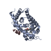
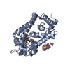
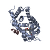
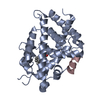
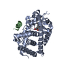
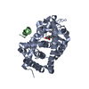
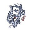
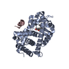
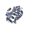
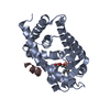
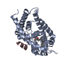
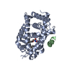
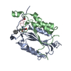
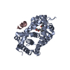
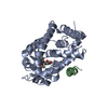
| Entry | Database: PDB / ID: 3vt8 | ||||||
|---|---|---|---|---|---|---|---|
| Title | Crystal structures of rat VDR-LBD with W282R mutation | ||||||
 Components Components |
| ||||||
 Keywords Keywords |  HORMONE RECEPTOR / Transcription HORMONE RECEPTOR / Transcription | ||||||
| Function / homology |  Function and homology information Function and homology informationnegative regulation of bone trabecula formation / Vitamin D (calciferol) metabolism / SUMOylation of intracellular receptors / Nuclear Receptor transcription pathway / bile acid nuclear receptor activity / response to bile acid / dense fibrillar component / positive regulation of parathyroid hormone secretion / phosphate ion transmembrane transport / apoptotic process involved in mammary gland involution ...negative regulation of bone trabecula formation / Vitamin D (calciferol) metabolism / SUMOylation of intracellular receptors / Nuclear Receptor transcription pathway / bile acid nuclear receptor activity / response to bile acid / dense fibrillar component / positive regulation of parathyroid hormone secretion / phosphate ion transmembrane transport / apoptotic process involved in mammary gland involution / cellular response to vitamin D / vitamin D binding / calcitriol binding / positive regulation of apoptotic process involved in mammary gland involution / vitamin D response element binding / lithocholic acid binding / positive regulation of keratinocyte differentiation / negative regulation of ossification / positive regulation of vitamin D receptor signaling pathway / vitamin D receptor signaling pathway / bile acid signaling pathway / intestinal absorption / response to aldosterone / mammary gland branching involved in pregnancy / regulation of calcium ion transport / decidualization / negative regulation of keratinocyte proliferation / heterochromatin / nuclear retinoid X receptor binding / T-tubule / lactation / skeletal system development / apoptotic signaling pathway / animal organ morphogenesis / euchromatin / mRNA transcription by RNA polymerase II / cell morphogenesis / nuclear matrix / response to calcium ion / intracellular calcium ion homeostasis / RNA polymerase II transcription regulator complex / cellular response to amyloid-beta / calcium ion transport / nuclear receptor activity / response to estradiol / heart development / sequence-specific DNA binding / cell differentiation / receptor complex / RNA polymerase II cis-regulatory region sequence-specific DNA binding / DNA-binding transcription factor activity / negative regulation of cell population proliferation / negative regulation of DNA-templated transcription / regulation of DNA-templated transcription / positive regulation of gene expression / regulation of transcription by RNA polymerase II / perinuclear region of cytoplasm / negative regulation of transcription by RNA polymerase II / positive regulation of transcription by RNA polymerase II / DNA binding / zinc ion binding / nucleusSimilarity search - Function | ||||||
| Biological species |  Rattus norvegicus (Norway rat) Rattus norvegicus (Norway rat) | ||||||
| Method | X-RAY DIFFRACTION / SYNCHROTRON / MOLECULAR REPLACEMENT / Resolution: 2.1 Å | ||||||
 Authors Authors | Nakabayashi, M. / Shimizu, M. / Ikura, T. / Ito, N. | ||||||
 Citation Citation | Journal: J.Med.Chem. / Year: 2013 Title: Crystal structures of hereditary vitamin D-resistant rickets-associated vitamin D receptor mutants R270L and W282R bound to 1,25-dihydroxyvitamin D3 and synthetic ligands. Authors: Nakabayashi, M. / Tsukahara, Y. / Iwasaki-Miyamoto, Y. / Mihori-Shimazaki, M. / Yamada, S. / Inaba, S. / Oda, M. / Shimizu, M. / Makishima, M. / Tokiwa, H. / Ikura, T. / Ito, N. | ||||||
| History |
|
- Structure visualization
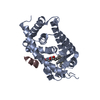
| Structure viewer | Molecule: MolmilJmol/JSmol |
|---|
- Downloads & links
-Download
| PDBx/mmCIF format | 3vt8.cif.gz | 66.8 KB | Display | PDBx/mmCIF format |
|---|---|---|---|---|
| PDB format | pdb3vt8.ent.gz | 47.5 KB | Display | PDB format |
| PDBx/mmJSON format | 3vt8.json.gz | Tree view | PDBx/mmJSON format | |
| Others |  Other downloads Other downloads |
-Validation report
| Arichive directory | https://data.pdbj.org/pub/pdb/validation_reports/vt/3vt8ftp://data.pdbj.org/pub/pdb/validation_reports/vt/3vt8 | HTTPS FTP |
|---|
-Related structure data
| Related structure data |  3vt3C  3vt4C  3vt5C  3vt6C 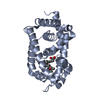 3vt7C  3vt9C  2zlcS S: Starting model for refinement C: citing same article ( |
|---|---|
| Similar structure data |


-Links
 PDBj
PDBj


- Assembly
| Deposited unit | 
| ||||||||
|---|---|---|---|---|---|---|---|---|---|
| 1 |
| ||||||||
| 2 | 
| ||||||||
| Unit cell |
|
-Components
| #1: Protein | Mass: 30566.021 Da / Num. of mol.: 1 / Fragment: UNP RESIDUES 116-423 / Mutation: W282R Source method: isolated from a genetically manipulated source Source: (gene. exp.) Rattus norvegicus (Norway rat) / Gene: Nr1i1, Vdr / Plasmid: pET14b / Production host:  Escherichia coli (E. coli) / Strain (production host): C41 / References: UniProt: P13053 Escherichia coli (E. coli) / Strain (production host): C41 / References: UniProt: P13053 |
|---|---|
| #2: Protein/peptide | Mass: 1570.898 Da / Num. of mol.: 1 / Source method: obtained synthetically / Details: PEPTIDE SYNTHESIS |
| #3: Chemical | ChemComp-YI3 / (  Mass: 460.732 Da / Num. of mol.: 1 / Source method: obtained synthetically / Formula: C30H52O3 Mass: 460.732 Da / Num. of mol.: 1 / Source method: obtained synthetically / Formula: C30H52O3 |
| #4: Water | ChemComp-HOH / Water Mass: 18.015 Da / Num. of mol.: 95 / Source method: isolated from a natural source / Formula: H2O Mass: 18.015 Da / Num. of mol.: 95 / Source method: isolated from a natural source / Formula: H2O |
-Experimental details
-Experiment
| Experiment | Method: X-RAY DIFFRACTION |
|---|
- Sample preparation
| Crystal | Density Matthews: 2.11 Å3/Da / Density % sol: 41.81 % |
|---|---|
| Crystal grow | Temperature: 293 K / Method: vapor diffusion, hanging drop / pH: 7 Details: MOPS-Na, di-Ammonium Citrate, PEG 4000, 2-propanol, pH 7.0, VAPOR DIFFUSION, HANGING DROP, temperature 293K |
-Data collection
| Diffraction source | Source: SYNCHROTRON / Site: Photon Factory  / Beamline: BL-6A / Wavelength: 0.978 Å / Beamline: BL-6A / Wavelength: 0.978 Å |
|---|---|
| Detector | Date: Feb 14, 2008 |
| Radiation | Protocol: SINGLE WAVELENGTH / Monochromatic (M) / Laue (L): M / Scattering type: x-ray |
| Radiation wavelength | Wavelength: 0.978 Å / Relative weight: 1 |
| Reflection | Resolution: 2.1→50 Å / Num. obs: 15047 / Biso Wilson estimate: 16.6 Å2 |
| Reflection shell | Resolution: 2.1→2.18 Å |
- Processing
| Software |
| ||||||||||||||||||||||||||||||||||||||||||||||||||||||||||||||||||||||||||||||||
|---|---|---|---|---|---|---|---|---|---|---|---|---|---|---|---|---|---|---|---|---|---|---|---|---|---|---|---|---|---|---|---|---|---|---|---|---|---|---|---|---|---|---|---|---|---|---|---|---|---|---|---|---|---|---|---|---|---|---|---|---|---|---|---|---|---|---|---|---|---|---|---|---|---|---|---|---|---|---|---|---|---|
| Refinement | Method to determine structure: MOLECULAR REPLACEMENT Starting model: 2ZLC Resolution: 2.1→38.26 Å / Rfactor Rfree error: 0.006 / Data cutoff high absF: 100897.41 / Data cutoff low absF: 0 / Isotropic thermal model: RESTRAINED / Cross valid method: THROUGHOUT / σ(F): 0
| ||||||||||||||||||||||||||||||||||||||||||||||||||||||||||||||||||||||||||||||||
| Solvent computation | Solvent model: FLAT MODEL / Bsol: 51.0914 Å2 / ksol: 0.363018 e/Å3 | ||||||||||||||||||||||||||||||||||||||||||||||||||||||||||||||||||||||||||||||||
| Displacement parameters | Biso mean: 33.8 Å2
| ||||||||||||||||||||||||||||||||||||||||||||||||||||||||||||||||||||||||||||||||
| Refine analyze |
| ||||||||||||||||||||||||||||||||||||||||||||||||||||||||||||||||||||||||||||||||
| Refinement step | Cycle: LAST / Resolution: 2.1→38.26 Å
| ||||||||||||||||||||||||||||||||||||||||||||||||||||||||||||||||||||||||||||||||
| Refine LS restraints |
| ||||||||||||||||||||||||||||||||||||||||||||||||||||||||||||||||||||||||||||||||
| LS refinement shell | Resolution: 2.1→2.23 Å / Rfactor Rfree error: 0.019 / Total num. of bins used: 6
| ||||||||||||||||||||||||||||||||||||||||||||||||||||||||||||||||||||||||||||||||
| Xplor file |
|
