 Movie
Movie Controller

[English] 日本語
 Yorodumi
- PDB-2hc4: Crystal structure of the LBD of VDR of Danio rerio in complex wit... -
+ Open data

- Basic information
| Entry | Database: PDB / ID: 2hc4 | ||||||
|---|---|---|---|---|---|---|---|
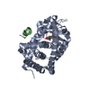
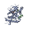
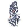
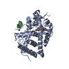
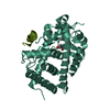
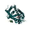
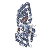
| Title | Crystal structure of the LBD of VDR of Danio rerio in complex with calcitriol | ||||||
 Components Components |
| ||||||
 Keywords Keywords |  GENE REGULATION / alpha helical sandwich GENE REGULATION / alpha helical sandwich | ||||||
| Function / homology |  Function and homology information Function and homology informationheart jogging / Vitamin D (calciferol) metabolism / : / SUMOylation of intracellular receptors / vitamin D binding / calcitriol binding / lithocholic acid binding / labyrinthine layer morphogenesis / regulation of thyroid hormone mediated signaling pathway / positive regulation of transcription from RNA polymerase II promoter by galactose ...heart jogging / Vitamin D (calciferol) metabolism / : / SUMOylation of intracellular receptors / vitamin D binding / calcitriol binding / lithocholic acid binding / labyrinthine layer morphogenesis / regulation of thyroid hormone mediated signaling pathway / positive regulation of transcription from RNA polymerase II promoter by galactose / positive regulation of female receptivity / hematopoietic stem cell proliferation / hypothalamus development / male mating behavior / NR1H2 & NR1H3 regulate gene expression to control bile acid homeostasis / heart looping / estrous cycle / cellular response to Thyroglobulin triiodothyronine / Synthesis of bile acids and bile salts / calcium ion homeostasis / Endogenous sterols / Synthesis of bile acids and bile salts via 27-hydroxycholesterol / Synthesis of bile acids and bile salts via 7alpha-hydroxycholesterol / nuclear retinoid X receptor binding / response to retinoic acid / histone acetyltransferase activity / regulation of cellular response to insulin stimulus / Recycling of bile acids and salts / histone acetyltransferase / cellular response to hormone stimulus / NR1H3 & NR1H2 regulate gene expression linked to cholesterol transport and efflux / positive regulation of adipose tissue development / peroxisome proliferator activated receptor signaling pathway / RORA activates gene expression / lactation / positive regulation of neuron differentiation / Regulation of lipid metabolism by PPARalpha / cerebellum development / ossification / BMAL1:CLOCK,NPAS2 activates circadian gene expression / SUMOylation of transcription cofactors / Activation of gene expression by SREBF (SREBP) / nuclear receptor coactivator activity / response to progesterone / nuclear estrogen receptor binding / nuclear receptor binding / hippocampus development / RNA polymerase II transcription regulatory region sequence-specific DNA binding / Heme signaling / mRNA transcription by RNA polymerase II / Transcriptional activation of mitochondrial biogenesis / PPARA activates gene expression / Cytoprotection by HMOX1 / cerebral cortex development / Transcriptional regulation of white adipocyte differentiation / RNA polymerase II transcription regulator complex / male gonad development / nuclear receptor activity / Circadian Clock / response to estradiol / HATs acetylate histones / Estrogen-dependent gene expression / transcription regulator complex / cell differentiation / transcription coactivator activity / protein dimerization activity / positive regulation of apoptotic process / RNA polymerase II cis-regulatory region sequence-specific DNA binding / DNA-templated transcription / chromatin binding / chromatin / regulation of DNA-templated transcription / positive regulation of DNA-templated transcription / negative regulation of transcription by RNA polymerase II / positive regulation of transcription by RNA polymerase II / protein-containing complex / zinc ion binding / nucleoplasm / nucleus / plasma membrane / cytosol / cytoplasmSimilarity search - Function | ||||||
| Biological species |  Danio rerio (zebrafish) Danio rerio (zebrafish) Homo sapiens (human) Homo sapiens (human) | ||||||
| Method | X-RAY DIFFRACTION / SYNCHROTRON / MOLECULAR REPLACEMENT / Resolution: 2.2 Å | ||||||
 Authors Authors | Ciesielski, F. / Rochel, N. / Moras, D. | ||||||
 Citation Citation | Journal: J.Steroid Biochem.Mol.Biol. / Year: 2007 Title: Adaptability of the Vitamin D nuclear receptor to the synthetic ligand Gemini: remodelling the LBP with one side chain rotation Authors: Ciesielski, F. / Rochel, N. / Moras, D. | ||||||
| History |
|
- Structure visualization
| Structure viewer | Molecule: MolmilJmol/JSmol |
|---|
- Downloads & links
-Download
| PDBx/mmCIF format | 2hc4.cif.gz | 67.8 KB | Display | PDBx/mmCIF format |
|---|---|---|---|---|
| PDB format | pdb2hc4.ent.gz | 48.3 KB | Display | PDB format |
| PDBx/mmJSON format | 2hc4.json.gz | Tree view | PDBx/mmJSON format | |
| Others |  Other downloads Other downloads |
-Validation report
| Arichive directory | https://data.pdbj.org/pub/pdb/validation_reports/hc/2hc4ftp://data.pdbj.org/pub/pdb/validation_reports/hc/2hc4 | HTTPS FTP |
|---|
-Related structure data
| Related structure data | 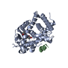 2hcdC 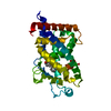 1db1S S: Starting model for refinement C: citing same article ( |
|---|---|
| Similar structure data |


-Links
 PDBj
PDBj


- Assembly
| Deposited unit | 
| ||||||||
|---|---|---|---|---|---|---|---|---|---|
| 1 | 
| ||||||||
| Unit cell |
|
-Components
| #1: Protein | Mass: 34060.672 Da / Num. of mol.: 1 / Fragment: Ligand binding domain Source method: isolated from a genetically manipulated source Source: (gene. exp.) Danio rerio (zebrafish) / Plasmid: pET28b / Species (production host): Escherichia coli / Production host:  Escherichia coli BL21(DE3) (bacteria) / Strain (production host): BL21DE3 / References: UniProt: Q9PTN2 Escherichia coli BL21(DE3) (bacteria) / Strain (production host): BL21DE3 / References: UniProt: Q9PTN2 |
|---|---|
| #2: Protein/peptide | Nuclear receptor coactivator 1 / NCoA-1 / Steroid receptor coactivator 1 / SRC-1 / RIP160 / Protein Hin-2 / NY-REN-52 antigen Mass: 1776.072 Da / Num. of mol.: 1 / Source method: obtained synthetically / Details: This sequence occurs naturally in humans / Source: (synth.) Homo sapiens (human) / References: UniProt: Q15788 |
| #3: Chemical | ChemComp-VDX / Calcitriol  Mass: 416.636 Da / Num. of mol.: 1 / Source method: obtained synthetically / Formula: C27H44O3 Mass: 416.636 Da / Num. of mol.: 1 / Source method: obtained synthetically / Formula: C27H44O3 |
| #4: Water | ChemComp-HOH / Water Mass: 18.015 Da / Num. of mol.: 94 / Source method: isolated from a natural source / Formula: H2O Mass: 18.015 Da / Num. of mol.: 94 / Source method: isolated from a natural source / Formula: H2O |
-Experimental details
-Experiment
| Experiment | Method: X-RAY DIFFRACTION / Number of used crystals: 1 |
|---|
- Sample preparation
| Crystal | Density Matthews: 2.31 Å3/Da / Density % sol: 46.77 % |
|---|---|
| Crystal grow | Temperature: 298 K / Method: vapor diffusion, hanging drop / pH: 6.5 Details: Li sulfate 1.6M, Mg sulfate 50mM, Bis Tris 0.1M, pH 6.5, VAPOR DIFFUSION, HANGING DROP, temperature 298K |
-Data collection
| Diffraction | Mean temperature: 100 K |
|---|---|
| Diffraction source | Source: SYNCHROTRON / Site: ESRF  / Beamline: BM30A / Wavelength: 0.9762 Å / Beamline: BM30A / Wavelength: 0.9762 Å |
| Detector | Type: MAR CCD 165 mm / Detector: CCD / Date: Sep 28, 2002 |
| Radiation | Monochromator: Si111 / Protocol: SINGLE WAVELENGTH / Monochromatic (M) / Laue (L): M / Scattering type: x-ray |
| Radiation wavelength | Wavelength: 0.9762 Å / Relative weight: 1 |
| Reflection | Resolution: 2.1→25 Å / Num. all: 20786 / Num. obs: 20786 / % possible obs: 98.9 % / Observed criterion σ(F): 1 / Observed criterion σ(I): 1 |
| Reflection shell | Resolution: 2.1→2.18 Å / % possible all: 99 |
- Processing
| Software |
| ||||||||||||||||||||
|---|---|---|---|---|---|---|---|---|---|---|---|---|---|---|---|---|---|---|---|---|---|
| Refinement | Method to determine structure: MOLECULAR REPLACEMENT Starting model: 1DB1 Resolution: 2.2→20 Å / σ(F): 2 / Stereochemistry target values: Engh & Huber
| ||||||||||||||||||||
| Refinement step | Cycle: LAST / Resolution: 2.2→20 Å
| ||||||||||||||||||||
| Refine LS restraints |
|
