 Movie
Movie Controller

[English] 日本語
 Yorodumi
- PDB-1rhk: Crystal structure of the complex of caspase-3 with a phenyl-propy... -
+ Open data

- Basic information
| Entry | Database: PDB / ID: 1rhk | ||||||
|---|---|---|---|---|---|---|---|
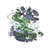
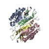
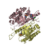
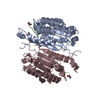
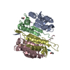
| Title | Crystal structure of the complex of caspase-3 with a phenyl-propyl-ketone inhibitor | ||||||
 Components Components |
| ||||||
 Keywords Keywords | HYDROLASE/HYDROLASE INHIBITOR /  CYSTEINE PROTEASE / CASPASE-3 / APOPAIN / CPP32 / YAMA / HYDROLASE-HYDROLASE INHIBITOR COMPLEX CYSTEINE PROTEASE / CASPASE-3 / APOPAIN / CPP32 / YAMA / HYDROLASE-HYDROLASE INHIBITOR COMPLEX | ||||||
| Function / homology |  Function and homology informationcaspase-3 / Stimulation of the cell death response by PAK-2p34 / phospholipase A2 activator activity / anterior neural tube closure / intrinsic apoptotic signaling pathway in response to osmotic stress / leukocyte apoptotic process / positive regulation of pyroptotic inflammatory response / glial cell apoptotic process / NADE modulates death signalling / luteolysis ...caspase-3 / Stimulation of the cell death response by PAK-2p34 / phospholipase A2 activator activity / anterior neural tube closure / intrinsic apoptotic signaling pathway in response to osmotic stress / leukocyte apoptotic process / positive regulation of pyroptotic inflammatory response / glial cell apoptotic process / NADE modulates death signalling / luteolysis / response to cobalt ion / cysteine-type endopeptidase activity involved in apoptotic signaling pathway / death-inducing signaling complex / cyclin-dependent protein serine/threonine kinase inhibitor activity / cellular response to staurosporine / Apoptosis induced DNA fragmentation / Apoptotic cleavage of cell adhesion proteins / cysteine-type endopeptidase activity involved in execution phase of apoptosis / Caspase activation via Dependence Receptors in the absence of ligand / death receptor binding / SMAC, XIAP-regulated apoptotic response / axonal fasciculation / Activation of caspases through apoptosome-mediated cleavage / Signaling by Hippo / SMAC (DIABLO) binds to IAPs / SMAC(DIABLO)-mediated dissociation of IAP:caspase complexes / cysteine-type endopeptidase activity involved in apoptotic process / fibroblast apoptotic process / execution phase of apoptosis / negative regulation of cytokine production / epithelial cell apoptotic process / platelet formation / Other interleukin signaling / positive regulation of amyloid-beta formation / pyroptotic inflammatory response / Apoptotic cleavage of cellular proteins / negative regulation of B cell proliferation / T cell homeostasis / negative regulation of activated T cell proliferation / neurotrophin TRK receptor signaling pathway / B cell homeostasis / protein maturation / negative regulation of cell cycle / response to X-ray / Caspase-mediated cleavage of cytoskeletal proteins / regulation of macroautophagy / response to amino acid / cell fate commitment / Pyroptosis / response to tumor necrosis factor / response to glucose / response to UV / response to glucocorticoid / keratinocyte differentiation / striated muscle cell differentiation / Degradation of the extracellular matrix / intrinsic apoptotic signaling pathway / erythrocyte differentiation / response to nicotine / apoptotic signaling pathway / hippocampus development / sensory perception of sound / protein catabolic process / regulation of protein stability / response to hydrogen peroxide / protein processing / neuron differentiation / response to wounding / positive regulation of neuron apoptotic process / response to estradiol / heart development / peptidase activity / neuron apoptotic process / protease binding / response to lipopolysaccharide / aspartic-type endopeptidase activity / response to hypoxia / learning or memory / response to xenobiotic stimulus / positive regulation of apoptotic process / cysteine-type endopeptidase activity / neuronal cell body / apoptotic process / DNA damage response / protein-containing complex binding / proteolysis / nucleoplasm / nucleus / cytosol / cytoplasm Function and homology informationcaspase-3 / Stimulation of the cell death response by PAK-2p34 / phospholipase A2 activator activity / anterior neural tube closure / intrinsic apoptotic signaling pathway in response to osmotic stress / leukocyte apoptotic process / positive regulation of pyroptotic inflammatory response / glial cell apoptotic process / NADE modulates death signalling / luteolysis ...caspase-3 / Stimulation of the cell death response by PAK-2p34 / phospholipase A2 activator activity / anterior neural tube closure / intrinsic apoptotic signaling pathway in response to osmotic stress / leukocyte apoptotic process / positive regulation of pyroptotic inflammatory response / glial cell apoptotic process / NADE modulates death signalling / luteolysis / response to cobalt ion / cysteine-type endopeptidase activity involved in apoptotic signaling pathway / death-inducing signaling complex / cyclin-dependent protein serine/threonine kinase inhibitor activity / cellular response to staurosporine / Apoptosis induced DNA fragmentation / Apoptotic cleavage of cell adhesion proteins / cysteine-type endopeptidase activity involved in execution phase of apoptosis / Caspase activation via Dependence Receptors in the absence of ligand / death receptor binding / SMAC, XIAP-regulated apoptotic response / axonal fasciculation / Activation of caspases through apoptosome-mediated cleavage / Signaling by Hippo / SMAC (DIABLO) binds to IAPs / SMAC(DIABLO)-mediated dissociation of IAP:caspase complexes / cysteine-type endopeptidase activity involved in apoptotic process / fibroblast apoptotic process / execution phase of apoptosis / negative regulation of cytokine production / epithelial cell apoptotic process / platelet formation / Other interleukin signaling / positive regulation of amyloid-beta formation / pyroptotic inflammatory response / Apoptotic cleavage of cellular proteins / negative regulation of B cell proliferation / T cell homeostasis / negative regulation of activated T cell proliferation / neurotrophin TRK receptor signaling pathway / B cell homeostasis / protein maturation / negative regulation of cell cycle / response to X-ray / Caspase-mediated cleavage of cytoskeletal proteins / regulation of macroautophagy / response to amino acid / cell fate commitment / Pyroptosis / response to tumor necrosis factor / response to glucose / response to UV / response to glucocorticoid / keratinocyte differentiation / striated muscle cell differentiation / Degradation of the extracellular matrix / intrinsic apoptotic signaling pathway / erythrocyte differentiation / response to nicotine / apoptotic signaling pathway / hippocampus development / sensory perception of sound / protein catabolic process / regulation of protein stability / response to hydrogen peroxide / protein processing / neuron differentiation / response to wounding / positive regulation of neuron apoptotic process / response to estradiol / heart development / peptidase activity / neuron apoptotic process / protease binding / response to lipopolysaccharide / aspartic-type endopeptidase activity / response to hypoxia / learning or memory / response to xenobiotic stimulus / positive regulation of apoptotic process / cysteine-type endopeptidase activity / neuronal cell body / apoptotic process / DNA damage response / protein-containing complex binding / proteolysis / nucleoplasm / nucleus / cytosol / cytoplasmSimilarity search - Function | ||||||
| Biological species |  Homo sapiens (human) Homo sapiens (human) | ||||||
| Method | X-RAY DIFFRACTION / MOLECULAR REPLACEMENT / Resolution: 2.5 Å | ||||||
 Authors Authors | Becker, J.W. / Rotonda, J. / Soisson, S.M. | ||||||
 Citation Citation | Journal: J.Med.Chem. / Year: 2004 Title: Reducing the Peptidyl Features of Caspase-3 Inhibitors: A Structural Analysis. Authors: Becker, J.W. / Rotonda, J. / Soisson, S.M. / Aspiotis, R. / Bayly, C. / Francoeur, S. / Gallant, M. / Garcia-Calvo, M. / Giroux, A. / Grimm, E. / Han, Y. / McKay, D. / Nicholson, D.W. / ...Authors: Becker, J.W. / Rotonda, J. / Soisson, S.M. / Aspiotis, R. / Bayly, C. / Francoeur, S. / Gallant, M. / Garcia-Calvo, M. / Giroux, A. / Grimm, E. / Han, Y. / McKay, D. / Nicholson, D.W. / Peterson, E. / Renaud, J. / Roy, S. / Thornberry, N. / Zamboni, R. | ||||||
| History |
|
- Structure visualization
| Structure viewer | Molecule: MolmilJmol/JSmol |
|---|
- Downloads & links
-Download
| PDBx/mmCIF format | 1rhk.cif.gz | 62.7 KB | Display | PDBx/mmCIF format |
|---|---|---|---|---|
| PDB format | pdb1rhk.ent.gz | 45 KB | Display | PDB format |
| PDBx/mmJSON format | 1rhk.json.gz | Tree view | PDBx/mmJSON format | |
| Others |  Other downloads Other downloads |
-Validation report
| Arichive directory | https://data.pdbj.org/pub/pdb/validation_reports/rh/1rhkftp://data.pdbj.org/pub/pdb/validation_reports/rh/1rhk | HTTPS FTP |
|---|
-Related structure data
| Related structure data | 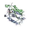 1re1C 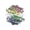 1rhjC 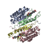 1rhmC  1rhqC 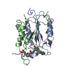 1rhrC  1rhuC 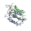 1pauS S: Starting model for refinement C: citing same article ( |
|---|---|
| Similar structure data |


-Links
 PDBj
PDBj


- Assembly
| Deposited unit | 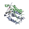
| ||||||||
|---|---|---|---|---|---|---|---|---|---|
| 1 | 
| ||||||||
| 2 | 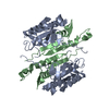
| ||||||||
| 3 | 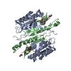
| ||||||||
| Unit cell |
| ||||||||
| Details | The second part of the biological assembly is generated by the two-fold axis: 1-x, y, -z |
-Components
| #1: Protein | Caspase 3 / Cysteine protease CPP32 / Yama protein / CPP-32 / Apopain / CASP-3 / SREBP cleavage activity 1 / SCA-1 Mass: 16639.902 Da / Num. of mol.: 1 / Fragment: P17 SUBUNIT Source method: isolated from a genetically manipulated source Source: (gene. exp.) Homo sapiens (human) / Gene: CASP3, CPP32 / Species (production host): Escherichia coli / Production host:  Escherichia coli BL21(DE3) (bacteria) / Strain (production host): BL21(DE3) Escherichia coli BL21(DE3) (bacteria) / Strain (production host): BL21(DE3)References: UniProt: P42574, Hydrolases; Acting on peptide bonds (peptidases); Cysteine endopeptidases |
|---|---|
| #2: Protein | Caspase 3 / Cysteine protease CPP32 / Yama protein / CPP-32 / Apopain / CASP-3 / SREBP cleavage activity 1 / SCA-1 Mass: 11910.604 Da / Num. of mol.: 1 / Fragment: P12 SUBUNIT Source method: isolated from a genetically manipulated source Source: (gene. exp.) Homo sapiens (human) / Gene: CASP3, CPP32 / Species (production host): Escherichia coli / Production host: Escherichia coli BL21(DE3) (bacteria) / Strain (production host): BL21(DE3)References: UniProt: P42574, Hydrolases; Acting on peptide bonds (peptidases); Cysteine endopeptidases |
| #3: Protein/peptide | 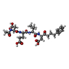  Type: Peptide-like / Class: Inhibitor / Mass: 604.649 Da / Num. of mol.: 1 / Source method: obtained synthetically / Details: The inhibitor is chemically synthesized. Type: Peptide-like / Class: Inhibitor / Mass: 604.649 Da / Num. of mol.: 1 / Source method: obtained synthetically / Details: The inhibitor is chemically synthesized.References: N-acetyl-L-alpha-aspartyl-L-alpha-glutamyl-N-[(1S)-1-(carboxymethyl)-2-oxo-5-phenylpentyl]-L-valinamide |
| #4: Water | ChemComp-HOH / Water Mass: 18.015 Da / Num. of mol.: 14 / Source method: isolated from a natural source / Formula: H2O Mass: 18.015 Da / Num. of mol.: 14 / Source method: isolated from a natural source / Formula: H2O |
-Experimental details
-Experiment
| Experiment | Method: X-RAY DIFFRACTION / Number of used crystals: 1 |
|---|
- Sample preparation
| Crystal | Density Matthews: 2.47 Å3/Da / Density % sol: 50.12 % |
|---|---|
| Crystal grow | Temperature: 293 K / Method: vapor diffusion, hanging drop / pH: 5.3 Details: 12% PEG-5000, 100 mM Citrate, 10 mM DTT, 3 mM NaN(3), pH 5.3, VAPOR DIFFUSION, HANGING DROP, temperature 293K |
-Data collection
| Diffraction | Mean temperature: 293 K |
|---|---|
| Diffraction source | Source: ROTATING ANODE / Type: RIGAKU RU200 / Wavelength: 1.5418 |
| Detector | Type: SIEMENS / Detector: AREA DETECTOR / Date: Apr 29, 1996 |
| Radiation | Monochromator: Graphite / Protocol: SINGLE WAVELENGTH / Monochromatic (M) / Laue (L): M / Scattering type: x-ray |
| Radiation wavelength | Wavelength: 1.5418 Å / Relative weight: 1 |
| Reflection | Resolution: 2.5→100 Å / Num. all: 10263 / Num. obs: 8371 / % possible obs: 81.6 % / Observed criterion σ(F): 0 / Observed criterion σ(I): 0 / Redundancy: 2.1 % / Biso Wilson estimate: 10.9 Å2 / Rmerge(I) obs: 0.08 / Net I/σ(I): 10.93 |
| Reflection shell | Resolution: 2.5→2.589 Å / Redundancy: 1.38 % / Rmerge(I) obs: 0.362 / Mean I/σ(I) obs: 2.3 / Num. unique all: 333 / % possible all: 33.3 |
- Processing
| Software |
| ||||||||||||||||||||||||||||||||||||
|---|---|---|---|---|---|---|---|---|---|---|---|---|---|---|---|---|---|---|---|---|---|---|---|---|---|---|---|---|---|---|---|---|---|---|---|---|---|
| Refinement | Method to determine structure: MOLECULAR REPLACEMENT Starting model: Protein part of 1pau.pdb Resolution: 2.5→19.98 Å / Rfactor Rfree error: 0.008 / Data cutoff high absF: 26033.7 / Data cutoff low absF: 0 / Isotropic thermal model: RESTRAINED / Cross valid method: THROUGHOUT / σ(F): 0 / σ(I): 0 / Stereochemistry target values: Engh & Huber / Details: BULK SOLVENT MODEL USED
| ||||||||||||||||||||||||||||||||||||
| Solvent computation | Solvent model: FLAT MODEL / Bsol: 12.987 Å2 / ksol: 0.299302 e/Å3 | ||||||||||||||||||||||||||||||||||||
| Displacement parameters | Biso mean: 19.5 Å2
| ||||||||||||||||||||||||||||||||||||
| Refine analyze |
| ||||||||||||||||||||||||||||||||||||
| Refinement step | Cycle: LAST / Resolution: 2.5→19.98 Å
| ||||||||||||||||||||||||||||||||||||
| Refine LS restraints |
| ||||||||||||||||||||||||||||||||||||
| LS refinement shell | Resolution: 2.5→2.66 Å / Rfactor Rfree error: 0.039 / Total num. of bins used: 6
| ||||||||||||||||||||||||||||||||||||
| Xplor file |
|
