 Movie
Movie Controller

[English] 日本語
 Yorodumi
- PDB-3vke: Contribution of the first K-homology domain of poly(C)-binding pr... -
+ Open data

- Basic information
| Entry | Database: PDB / ID: 3vke | ||||||
|---|---|---|---|---|---|---|---|
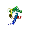
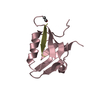
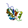
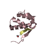
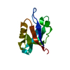
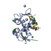
| Title | Contribution of the first K-homology domain of poly(C)-binding protein 1 to its affinity and specificity for C-rich oligonucleotides | ||||||
 Components Components |
| ||||||
 Keywords Keywords | DNA BINDING PROTEIN/DNA / PCBP-1 / DNA RNA BINDING PROTEIN /  KH DOMAIN / DNA BINDING PROTEIN-DNA complex KH DOMAIN / DNA BINDING PROTEIN-DNA complex | ||||||
| Function / homology |  Function and homology information Function and homology informationsequence-specific single stranded DNA binding / Processing of Capped Intron-Containing Pre-mRNA / mRNA Splicing - Major Pathway / cytoplasmic ribonucleoprotein granule / single-stranded DNA binding / postsynaptic density / DNA-binding transcription factor activity, RNA polymerase II-specific / nuclear speck / cadherin binding / ribonucleoprotein complex ...sequence-specific single stranded DNA binding / Processing of Capped Intron-Containing Pre-mRNA / mRNA Splicing - Major Pathway / cytoplasmic ribonucleoprotein granule / single-stranded DNA binding / postsynaptic density / DNA-binding transcription factor activity, RNA polymerase II-specific / nuclear speck / cadherin binding / ribonucleoprotein complex / viral RNA genome replication / mRNA binding / positive regulation of transcription by RNA polymerase II / RNA binding / extracellular exosome / nucleoplasm / membrane / nucleus / cytosol / cytoplasmSimilarity search - Function | ||||||
| Biological species |  Homo sapiens (human) Homo sapiens (human) | ||||||
| Method | X-RAY DIFFRACTION / SYNCHROTRON / MOLECULAR REPLACEMENT / Resolution: 1.77 Å | ||||||
 Authors Authors | Traore, D.A.K. / Wilce, M.C.J. / Wilce, J.A. | ||||||
 Citation Citation | Journal: Nucleic Acids Res. / Year: 2012 Title: Contribution of the first K-homology domain of poly(C)-binding protein 1 to its affinity and specificity for C-rich oligonucleotides Authors: Yoga, Y.M.K. / Traore, D.A.K. / Sidiqi, M. / Szeto, C. / Pendini, N.R. / Barker, A. / Leedman, P.J. / Wilce, J.A. / Wilce, M.C.J. | ||||||
| History |
|
- Structure visualization
| Structure viewer | Molecule: MolmilJmol/JSmol |
|---|
- Downloads & links
-Download
| PDBx/mmCIF format | 3vke.cif.gz | 147.8 KB | Display | PDBx/mmCIF format |
|---|---|---|---|---|
| PDB format | pdb3vke.ent.gz | 116.6 KB | Display | PDB format |
| PDBx/mmJSON format | 3vke.json.gz | Tree view | PDBx/mmJSON format | |
| Others |  Other downloads Other downloads |
-Validation report
| Arichive directory | https://data.pdbj.org/pub/pdb/validation_reports/vk/3vkeftp://data.pdbj.org/pub/pdb/validation_reports/vk/3vke | HTTPS FTP |
|---|
-Related structure data
| Related structure data | 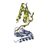 1ztgSC S: Starting model for refinement C: citing same article ( |
|---|---|
| Similar structure data |


-Links
 PDBj
PDBj


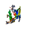
- Assembly
| Deposited unit | 
| ||||||||
|---|---|---|---|---|---|---|---|---|---|
| 1 | 
| ||||||||
| 2 | 
| ||||||||
| 3 | 
| ||||||||
| 4 | 
| ||||||||
| Unit cell |
|
-Components
| #1: Protein | Mass: 8462.802 Da / Num. of mol.: 4 / Fragment: KH1 domain / Mutation: C54S Source method: isolated from a genetically manipulated source Source: (gene. exp.) Homo sapiens (human) / Gene: PCBP1 / Plasmid: pET15b / Production host:  Escherichia coli (E. coli) / Strain (production host): BL21(DE3) / References: UniProt: Q15365 Escherichia coli (E. coli) / Strain (production host): BL21(DE3) / References: UniProt: Q15365#2: DNA chain | Mass: 1738.183 Da / Num. of mol.: 4 / Source method: obtained synthetically #3: Water | ChemComp-HOH / | Water Mass: 18.015 Da / Num. of mol.: 71 / Source method: isolated from a natural source / Formula: H2O Mass: 18.015 Da / Num. of mol.: 71 / Source method: isolated from a natural source / Formula: H2O |
|---|
-Experimental details
-Experiment
| Experiment | Method: X-RAY DIFFRACTION / Number of used crystals: 1 |
|---|
- Sample preparation
| Crystal | Density Matthews: 2.35 Å3/Da / Density % sol: 47.77 % |
|---|---|
| Crystal grow | Temperature: 293 K / Method: vapor diffusion, hanging drop / pH: 4.2 Details: 0.1M phosphate-citrate, 40%(v/v) PEG 300, pH 4.2, VAPOR DIFFUSION, HANGING DROP, temperature 293K |
-Data collection
| Diffraction | Mean temperature: 100 K |
|---|---|
| Diffraction source | Source: SYNCHROTRON / Site: Australian Synchrotron  / Beamline: MX1 / Wavelength: 0.9801 Å / Beamline: MX1 / Wavelength: 0.9801 Å |
| Detector | Type: ADSC QUANTUM 315r / Detector: CCD / Date: Mar 5, 2010 |
| Radiation | Protocol: SINGLE WAVELENGTH / Monochromatic (M) / Laue (L): M / Scattering type: x-ray |
| Radiation wavelength | Wavelength: 0.9801 Å / Relative weight: 1 |
| Reflection | Resolution: 1.77→38.51 Å / Num. obs: 36143 / Observed criterion σ(I): 3 / Redundancy: 3.2 % / Rmerge(I) obs: 0.09 / Net I/σ(I): 7.9 |
- Processing
| Software |
| ||||||||||||||||||||||||||||||||||||||||||||||||||||||||||||||||||||||
|---|---|---|---|---|---|---|---|---|---|---|---|---|---|---|---|---|---|---|---|---|---|---|---|---|---|---|---|---|---|---|---|---|---|---|---|---|---|---|---|---|---|---|---|---|---|---|---|---|---|---|---|---|---|---|---|---|---|---|---|---|---|---|---|---|---|---|---|---|---|---|---|
| Refinement | Method to determine structure: MOLECULAR REPLACEMENT Starting model: 1ZTG Resolution: 1.77→27.16 Å / Cor.coef. Fo:Fc: 0.963 / Cor.coef. Fo:Fc free: 0.944 / Occupancy max: 1 / Occupancy min: 0 / SU B: 4.484 / SU ML: 0.065 / Cross valid method: THROUGHOUT / σ(F): 0 / ESU R: 0.145 / ESU R Free: 0.107 / Stereochemistry target values: MAXIMUM LIKELIHOOD Details: HYDROGENS HAVE BEEN ADDED IN THE RIDING POSITIONS U VALUES: REFINED INDIVIDUALLY
| ||||||||||||||||||||||||||||||||||||||||||||||||||||||||||||||||||||||
| Solvent computation | Ion probe radii: 0.8 Å / Shrinkage radii: 0.8 Å / VDW probe radii: 1.4 Å / Solvent model: MASK | ||||||||||||||||||||||||||||||||||||||||||||||||||||||||||||||||||||||
| Displacement parameters | Biso max: 90.59 Å2 / Biso mean: 31.5288 Å2 / Biso min: 13.06 Å2
| ||||||||||||||||||||||||||||||||||||||||||||||||||||||||||||||||||||||
| Refinement step | Cycle: LAST / Resolution: 1.77→27.16 Å
| ||||||||||||||||||||||||||||||||||||||||||||||||||||||||||||||||||||||
| Refine LS restraints |
| ||||||||||||||||||||||||||||||||||||||||||||||||||||||||||||||||||||||
| LS refinement shell | Resolution: 1.77→1.816 Å / Total num. of bins used: 20
|
